


Crystal structure of the PNT domain of human ETS2
Newman, J.A., Cooper, C.D.O., Krojer, T., Shrestha, L., Burgess-brown, N., Arrowsmith, C.H., Bountra, C., Edwards, A., Gileadi, O.To be published.
Experimental Data Snapshot
wwPDB Validation 3D Report Full Report

Entity ID: 1 | |||||
|---|---|---|---|---|---|
| Molecule | Chains | Sequence Length | Organism | Details | Image |
| Protein C-ets-2 | 97 | Homo sapiens | Mutation(s): 0 Gene Names: ETS2 | 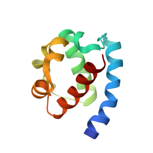 | |
UniProt & NIH Common Fund Data Resources | |||||
Find proteins for P15036 (Homo sapiens) Explore P15036 Go to UniProtKB: P15036 | |||||
PHAROS: P15036 GTEx: ENSG00000157557 | |||||
Entity Groups | |||||
| Sequence Clusters | 30% Identity50% Identity70% Identity90% Identity95% Identity100% Identity | ||||
| UniProt Group | P15036 | ||||
Sequence AnnotationsExpand | |||||
| |||||
| Ligands 3 Unique | |||||
|---|---|---|---|---|---|
| ID | Chains | Name / Formula / InChI Key | 2D Diagram | 3D Interactions | |
| GOL Query on GOL | D [auth A], F [auth B], G [auth B] | GLYCEROL C3 H8 O3 PEDCQBHIVMGVHV-UHFFFAOYSA-N |  | ||
| ACT Query on ACT | E [auth A], H [auth B] | ACETATE ION C2 H3 O2 QTBSBXVTEAMEQO-UHFFFAOYSA-M |  | ||
| CA Query on CA | C [auth A] | CALCIUM ION Ca BHPQYMZQTOCNFJ-UHFFFAOYSA-N |  | ||
| Length ( Å ) | Angle ( ˚ ) |
|---|---|
| a = 64.44 | α = 90 |
| b = 64.44 | β = 90 |
| c = 263.71 | γ = 120 |
| Software Name | Purpose |
|---|---|
| ADSC | data collection |
| SHARP | phasing |
| BUSTER | refinement |
| XDS | data reduction |
| XDS | data scaling |
RCSB PDB (citation) is hosted by
RCSB PDB is a member of the

