4LRH

Crystal structure of human folate receptor alpha in complex with folic acid


Experimental Data Snapshot

  • Method: X-RAY DIFFRACTION
  • Resolution: 2.80 Å
  • R-Value Free: 0.267 
  • R-Value Work: 0.227 

wwPDB Validation   3D Report Full Report


Ligand Structure Quality Assessment 


This is version 1.3 of the entry. See complete history


Literature

Structural basis for molecular recognition of folic acid by folate receptors.

Chen, C.Ke, J.Zhou, X.E.Yi, W.Brunzelle, J.S.Li, J.Yong, E.L.Xu, H.E.Melcher, K.

(2013) Nature 500: 486-489

  • DOI: https://doi.org/10.1038/nature12327
  • Primary Citation of Related Structures:  
    4LRH

  • PubMed Abstract: 

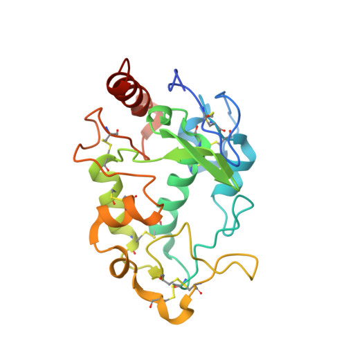
    Folate receptors (FRα, FRβ and FRγ) are cysteine-rich cell-surface glycoproteins that bind folate with high affinity to mediate cellular uptake of folate. Although expressed at very low levels in most tissues, folate receptors, especially FRα, are expressed at high levels in numerous cancers to meet the folate demand of rapidly dividing cells under low folate conditions. The folate dependency of many tumours has been therapeutically and diagnostically exploited by administration of anti-FRα antibodies, high-affinity antifolates, folate-based imaging agents and folate-conjugated drugs and toxins. To understand how folate binds its receptors, we determined the crystal structure of human FRα in complex with folic acid at 2.8 Å resolution. FRα has a globular structure stabilized by eight disulphide bonds and contains a deep open folate-binding pocket comprised of residues that are conserved in all receptor subtypes. The folate pteroate moiety is buried inside the receptor, whereas its glutamate moiety is solvent-exposed and sticks out of the pocket entrance, allowing it to be conjugated to drugs without adversely affecting FRα binding. The extensive interactions between the receptor and ligand readily explain the high folate-binding affinity of folate receptors and provide a template for designing more specific drugs targeting the folate receptor system.


  • Organizational Affiliation

    Program for Structural Biology and Drug Discovery, Van Andel Research Institute, 333 Bostwick Avenue North East, Grand Rapids, Michigan 49503, USA.


Macromolecules
Find similar proteins by:  (by identity cutoff)  |  3D Structure
Entity ID: 1
MoleculeChains Sequence LengthOrganismDetailsImage
Folate receptor alpha
A, B, C, D, E
A, B, C, D, E, F, G, H
218Homo sapiensMutation(s): 0 
Gene Names: FOLR1FOLR
UniProt & NIH Common Fund Data Resources
Find proteins for P15328 (Homo sapiens)
Explore P15328 
Go to UniProtKB:  P15328
PHAROS:  P15328
GTEx:  ENSG00000110195 
Entity Groups  
Sequence Clusters30% Identity50% Identity70% Identity90% Identity95% Identity100% Identity
UniProt GroupP15328
Sequence Annotations
Expand
  • Reference Sequence
Small Molecules
Ligands 2 Unique
IDChains Name / Formula / InChI Key2D Diagram3D Interactions
FOL
Query on FOL

Download Ideal Coordinates CCD File 
BA [auth F]
FA [auth G]
I [auth A]
IA [auth H]
M [auth B]
BA [auth F],
FA [auth G],
I [auth A],
IA [auth H],
M [auth B],
Q [auth C],
T [auth D],
X [auth E]
FOLIC ACID
C19 H19 N7 O6
OVBPIULPVIDEAO-LBPRGKRZSA-N
NAG
Query on NAG

Download Ideal Coordinates CCD File 
AA [auth E]
CA [auth F]
DA [auth F]
EA [auth F]
GA [auth G]
AA [auth E],
CA [auth F],
DA [auth F],
EA [auth F],
GA [auth G],
HA [auth G],
J [auth A],
JA [auth H],
K [auth A],
KA [auth H],
L [auth A],
LA [auth H],
N [auth B],
O [auth B],
P [auth B],
R [auth C],
S [auth C],
U [auth D],
V [auth D],
W [auth D],
Y [auth E],
Z [auth E]
2-acetamido-2-deoxy-beta-D-glucopyranose
C8 H15 N O6
OVRNDRQMDRJTHS-FMDGEEDCSA-N
Binding Affinity Annotations 
IDSourceBinding Affinity
FOL PDBBind:  4LRH Kd: 0.19 (nM) from 1 assay(s)
Experimental Data & Validation

Experimental Data

  • Method: X-RAY DIFFRACTION
  • Resolution: 2.80 Å
  • R-Value Free: 0.267 
  • R-Value Work: 0.227 
  • Space Group: P 21 21 21
Unit Cell:
Length ( Å )Angle ( ˚ )
a = 90.021α = 90
b = 144.589β = 90
c = 216.839γ = 90
Software Package:
Software NamePurpose
XDSdata scaling
SHELXSphasing
REFMACrefinement
XDSdata reduction
SCALAdata scaling

Structure Validation

View Full Validation Report


Ligand Structure Quality Assessment 


Entry History 

Deposition Data

Revision History  (Full details and data files)

  • Version 1.0: 2013-07-31
    Type: Initial release
  • Version 1.1: 2013-09-04
    Changes: Database references
  • Version 1.2: 2015-04-08
    Changes: Structure summary
  • Version 1.3: 2020-07-29
    Type: Remediation
    Reason: Carbohydrate remediation
    Changes: Data collection, Database references, Derived calculations, Structure summary