4KXH

The X-ray crystal structure of a dimeric variant of human pancreatic ribonuclease


Experimental Data Snapshot

  • Method: X-RAY DIFFRACTION
  • Resolution: 2.70 Å
  • R-Value Free: 0.246 
  • R-Value Work: 0.198 
  • R-Value Observed: 0.201 

wwPDB Validation   3D Report Full Report


This is version 1.2 of the entry. See complete history


Literature

Three-dimensional domain swapping and supramolecular protein assembly: insights from the X-ray structure of a dimeric swapped variant of human pancreatic RNase.

Pica, A.Merlino, A.Buell, A.K.Knowles, T.P.Pizzo, E.D'Alessio, G.Sica, F.Mazzarella, L.

(2013) Acta Crystallogr D Biol Crystallogr 69: 2116-2123

  • DOI: https://doi.org/10.1107/S0907444913020507
  • Primary Citation of Related Structures:  
    4KXH

  • PubMed Abstract: 

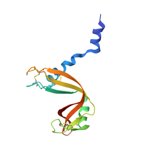
    The deletion of five residues in the loop connecting the N-terminal helix to the core of monomeric human pancreatic ribonuclease leads to the formation of an enzymatically active domain-swapped dimer (desHP). The crystal structure of desHP reveals the generation of an intriguing fibril-like aggregate of desHP molecules that extends along the c crystallographic axis. Dimers are formed by three-dimensional domain swapping. Tetramers are formed by the aggregation of swapped dimers with slightly different quaternary structures. The tetramers interact in such a way as to form an infinite rod-like structure that propagates throughout the crystal. The observed supramolecular assembly captured in the crystal predicts that desHP fibrils could form in solution; this has been confirmed by atomic force microscopy. These results provide new evidence that three-dimensional domain swapping can be a mechanism for the formation of elaborate large assemblies in which the protein, apart from the swapping, retains its original fold.


  • Organizational Affiliation

    Department of Chemical Sciences, University of Naples `Federico II', Via Cintia, 80126 Naples, Italy.


Macromolecules
Find similar proteins by:  (by identity cutoff)  |  3D Structure
Entity ID: 1
MoleculeChains Sequence LengthOrganismDetailsImage
Ribonuclease pancreatic
A, B, C, D
123Homo sapiensMutation(s): 0 
Gene Names: RNASE1RIB1RNS1
EC: 3.1.27.5
UniProt & NIH Common Fund Data Resources
Find proteins for P07998 (Homo sapiens)
Explore P07998 
Go to UniProtKB:  P07998
PHAROS:  P07998
GTEx:  ENSG00000129538 
Entity Groups  
Sequence Clusters30% Identity50% Identity70% Identity90% Identity95% Identity100% Identity
UniProt GroupP07998
Sequence Annotations
Expand
  • Reference Sequence
Small Molecules
Ligands 3 Unique
IDChains Name / Formula / InChI Key2D Diagram3D Interactions
SO4
Query on SO4

Download Ideal Coordinates CCD File 
E [auth A]
F [auth A]
G [auth B]
H [auth B]
I [auth B]
E [auth A],
F [auth A],
G [auth B],
H [auth B],
I [auth B],
J [auth B],
L [auth C],
M [auth C],
N [auth C],
O [auth C],
P [auth C],
S [auth D],
T [auth D]
SULFATE ION
O4 S
QAOWNCQODCNURD-UHFFFAOYSA-L
CL
Query on CL

Download Ideal Coordinates CCD File 
R [auth C]CHLORIDE ION
Cl
VEXZGXHMUGYJMC-UHFFFAOYSA-M
NA
Query on NA

Download Ideal Coordinates CCD File 
K [auth B],
Q [auth C],
U [auth D]
SODIUM ION
Na
FKNQFGJONOIPTF-UHFFFAOYSA-N
Experimental Data & Validation

Experimental Data

  • Method: X-RAY DIFFRACTION
  • Resolution: 2.70 Å
  • R-Value Free: 0.246 
  • R-Value Work: 0.198 
  • R-Value Observed: 0.201 
  • Space Group: P 21 21 21
Unit Cell:
Length ( Å )Angle ( ˚ )
a = 71.506α = 90
b = 74.319β = 90
c = 128.533γ = 90
Software Package:
Software NamePurpose
CCP4refinement
PHASERphasing
REFMACrefinement
HKL-2000data reduction
HKL-2000data scaling

Structure Validation

View Full Validation Report


Entry History 

Deposition Data

Revision History  (Full details and data files)

  • Version 1.0: 2013-10-02
    Type: Initial release
  • Version 1.1: 2014-09-24
    Changes: Database references
  • Version 1.2: 2017-11-15
    Changes: Refinement description