Membrane Curvature Protein Exhibits Interdomain Flexibility and Binds a Small GTPase.
King, G.J., Stockli, J., Hu, S.H., Winnen, B., Duprez, W.G., Meoli, C.C., Junutula, J.R., Jarrott, R.J., James, D.E., Whitten, A.E., Martin, J.L.(2012) J Biol Chem 287: 40996-41006
- PubMed: 23055524
- DOI: https://doi.org/10.1074/jbc.M112.349803
- Primary Citation of Related Structures:
4H8S - PubMed Abstract:
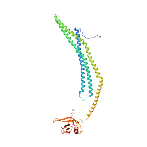
The APPL1 and APPL2 proteins (APPL (adaptor protein, phosphotyrosine interaction, pleckstrin homology (PH) domain, and leucine zipper-containing protein)) are localized to their own endosomal subcompartment and interact with a wide range of proteins and small molecules at the cell surface and in the nucleus. They play important roles in signal transduction through their ability to act as Rab effectors. (Rabs are a family of Ras GTPases involved in membrane trafficking.) Both APPL1 and APPL2 comprise an N-terminal membrane-curving BAR (Bin-amphiphysin-Rvs) domain linked to a PH domain and a C-terminal phosphotyrosine-binding domain. The structure and interactions of APPL1 are well characterized, but little is known about APPL2. Here, we report the crystal structure and low resolution solution structure of the BARPH domains of APPL2. We identify a previously undetected hinge site for rotation between the two domains and speculate that this motion may regulate APPL2 functions. We also identified Rab binding partners of APPL2 and show that these differ from those of APPL1, suggesting that APPL-Rab interaction partners have co-evolved over time. Isothermal titration calorimetry data reveal the interaction between APPL2 and Rab31 has a K(d) of 140 nM. Together with other biophysical data, we conclude the stoichiometry of the complex is 2:2.
Organizational Affiliation:
Institute for Molecular Bioscience, University of Queensland, Brisbane, Queensland 4072, Australia.