4GOV

The crystal structure of human fascin 1 S39D mutant


Experimental Data Snapshot

  • Method: X-RAY DIFFRACTION
  • Resolution: 2.20 Å
  • R-Value Free: 0.234 
  • R-Value Work: 0.196 
  • R-Value Observed: 0.198 

wwPDB Validation   3D Report Full Report


This is version 1.3 of the entry. See complete history


Literature

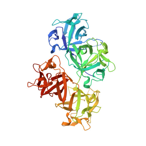
Molecular mechanism of fascin function in filopodial formation.

Yang, S.Huang, F.K.Huang, J.Chen, S.Jakoncic, J.Leo-Macias, A.Diaz-Avalos, R.Chen, L.Zhang, J.J.Huang, X.Y.

(2013) J Biol Chem 288: 274-284

  • DOI: https://doi.org/10.1074/jbc.M112.427971
  • Primary Citation of Related Structures:  
    4GOV, 4GOY, 4GP0, 4GP3

  • PubMed Abstract: 

    Filopodia are cell surface protrusions that are essential for cell migration. This finger-like structure is supported by rigid tightly bundled actin filaments. The protein responsible for actin bundling in filopodia is fascin. However, the mechanism by which fascin functions in filopodial formation is not clear. Here we provide biochemical, cryo-electron tomographic, and x-ray crystal structural data demonstrating the unique structural characteristics of fascin. Systematic mutagenesis studies on 100 mutants of fascin indicate that there are two major actin-binding sites on fascin. Crystal structures of four fascin mutants reveal concerted conformational changes in fascin from inactive to active states in the process of actin bundling. Mutations in any one of the actin-binding sites impair the cellular function of fascin in filopodial formation. Altogether, our data reveal the molecular mechanism of fascin function in filopodial formation.


  • Organizational Affiliation

    Department of Physiology, Cornell University Weill Medical College, New York, New York 10065, USA.


Macromolecules
Find similar proteins by:  (by identity cutoff)  |  3D Structure
Entity ID: 1
MoleculeChains Sequence LengthOrganismDetailsImage
Fascin
A, B
493Homo sapiensMutation(s): 1 
Gene Names: FAN1FSCN1HSNSNL
UniProt & NIH Common Fund Data Resources
Find proteins for Q16658 (Homo sapiens)
Explore Q16658 
Go to UniProtKB:  Q16658
PHAROS:  Q16658
GTEx:  ENSG00000075618 
Entity Groups  
Sequence Clusters30% Identity50% Identity70% Identity90% Identity95% Identity100% Identity
UniProt GroupQ16658
Sequence Annotations
Expand
  • Reference Sequence
Small Molecules
Ligands 4 Unique
IDChains Name / Formula / InChI Key2D Diagram3D Interactions
DTT
Query on DTT

Download Ideal Coordinates CCD File 
M [auth A]2,3-DIHYDROXY-1,4-DITHIOBUTANE
C4 H10 O2 S2
VHJLVAABSRFDPM-IMJSIDKUSA-N
GOL
Query on GOL

Download Ideal Coordinates CCD File 
AA [auth B]
BA [auth B]
CA [auth B]
DA [auth B]
EA [auth B]
AA [auth B],
BA [auth B],
CA [auth B],
DA [auth B],
EA [auth B],
FA [auth B],
J [auth A],
K [auth A],
L [auth A],
Z [auth B]
GLYCEROL
C3 H8 O3
PEDCQBHIVMGVHV-UHFFFAOYSA-N
BR
Query on BR

Download Ideal Coordinates CCD File 
C [auth A]
D [auth A]
N [auth B]
O [auth B]
P [auth B]
C [auth A],
D [auth A],
N [auth B],
O [auth B],
P [auth B],
Q [auth B]
BROMIDE ION
Br
CPELXLSAUQHCOX-UHFFFAOYSA-M
CL
Query on CL

Download Ideal Coordinates CCD File 
E [auth A]
F [auth A]
G [auth A]
H [auth A]
I [auth A]
E [auth A],
F [auth A],
G [auth A],
H [auth A],
I [auth A],
R [auth B],
S [auth B],
T [auth B],
U [auth B],
V [auth B],
W [auth B],
X [auth B],
Y [auth B]
CHLORIDE ION
Cl
VEXZGXHMUGYJMC-UHFFFAOYSA-M
Experimental Data & Validation

Experimental Data

  • Method: X-RAY DIFFRACTION
  • Resolution: 2.20 Å
  • R-Value Free: 0.234 
  • R-Value Work: 0.196 
  • R-Value Observed: 0.198 
  • Space Group: C 1 2 1
Unit Cell:
Length ( Å )Angle ( ˚ )
a = 161.25α = 90
b = 70.723β = 130.68
c = 111.058γ = 90
Software Package:
Software NamePurpose
HKL-2000data collection
PHASERphasing
REFMACrefinement
HKL-2000data reduction
SCALEPACKdata scaling

Structure Validation

View Full Validation Report


Entry History 

Deposition Data

Revision History  (Full details and data files)

  • Version 1.0: 2012-11-28
    Type: Initial release
  • Version 1.1: 2012-12-26
    Changes: Database references
  • Version 1.2: 2013-01-30
    Changes: Database references
  • Version 1.3: 2023-09-13
    Changes: Data collection, Database references, Derived calculations, Refinement description