4GGF

Crystal structure of Mn2+ bound calprotectin


Experimental Data Snapshot

  • Method: X-RAY DIFFRACTION
  • Resolution: 1.60 Å
  • R-Value Free: 0.202 
  • R-Value Work: 0.177 
  • R-Value Observed: 0.178 

wwPDB Validation   3D Report Full Report


This is version 1.3 of the entry. See complete history


Literature

Molecular basis for manganese sequestration by calprotectin and roles in the innate immune response to invading bacterial pathogens.

Damo, S.M.Kehl-Fie, T.E.Sugitani, N.Holt, M.E.Rathi, S.Murphy, W.J.Zhang, Y.Betz, C.Hench, L.Fritz, G.Skaar, E.P.Chazin, W.J.

(2013) Proc Natl Acad Sci U S A 110: 3841-3846

  • DOI: https://doi.org/10.1073/pnas.1220341110
  • Primary Citation of Related Structures:  
    4GGF

  • PubMed Abstract: 

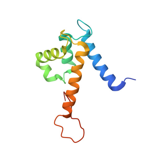
    The S100A8/S100A9 heterodimer calprotectin (CP) functions in the host response to pathogens through a mechanism termed "nutritional immunity." CP binds Mn(2+) and Zn(2+) with high affinity and starves bacteria of these essential nutrients. Combining biophysical, structural, and microbiological analysis, we identified the molecular basis of Mn(2+) sequestration. The asymmetry of the CP heterodimer creates a single Mn(2+)-binding site from six histidine residues, which distinguishes CP from all other Mn(2+)-binding proteins. Analysis of CP mutants with altered metal-binding properties revealed that, despite both Mn(2+) and Zn(2+) being essential metals, maximal growth inhibition of multiple bacterial pathogens requires Mn(2+) sequestration. These data establish the importance of Mn(2+) sequestration in defense against infection, explain the broad-spectrum antimicrobial activity of CP relative to other S100 proteins, and clarify the impact of metal depletion on the innate immune response to infection.


  • Organizational Affiliation

    Department of Biochemistry, and Center for Structural Biology, Vanderbilt University, Nashville, TN 37232-8725, USA.


Macromolecules
Find similar proteins by:  (by identity cutoff)  |  3D Structure
Entity ID: 1
MoleculeChains Sequence LengthOrganismDetailsImage
Protein S100-A8A,
C [auth K],
E [auth S],
G [auth U]
93Homo sapiensMutation(s): 0 
Gene Names: S100A8CAGACFAGMRP8
UniProt & NIH Common Fund Data Resources
Find proteins for P05109 (Homo sapiens)
Explore P05109 
Go to UniProtKB:  P05109
PHAROS:  P05109
GTEx:  ENSG00000143546 
Entity Groups  
Sequence Clusters30% Identity50% Identity70% Identity90% Identity95% Identity100% Identity
UniProt GroupP05109
Sequence Annotations
Expand
  • Reference Sequence
Find similar proteins by:  (by identity cutoff)  |  3D Structure
Entity ID: 2
MoleculeChains Sequence LengthOrganismDetailsImage
Protein S100-A9B [auth C],
D [auth L],
F [auth T],
H [auth V]
114Homo sapiensMutation(s): 0 
Gene Names: S100A9CAGBCFAGMRP14
UniProt & NIH Common Fund Data Resources
Find proteins for P06702 (Homo sapiens)
Explore P06702 
Go to UniProtKB:  P06702
PHAROS:  P06702
GTEx:  ENSG00000163220 
Entity Groups  
Sequence Clusters30% Identity50% Identity70% Identity90% Identity95% Identity100% Identity
UniProt GroupP06702
Sequence Annotations
Expand
  • Reference Sequence
Small Molecules
Ligands 4 Unique
IDChains Name / Formula / InChI Key2D Diagram3D Interactions
SO4
Query on SO4

Download Ideal Coordinates CCD File 
HA [auth V],
O [auth K],
T [auth L]
SULFATE ION
O4 S
QAOWNCQODCNURD-UHFFFAOYSA-L
GOL
Query on GOL

Download Ideal Coordinates CCD File 
DA [auth T]
EA [auth T]
N [auth C]
Q [auth K]
X [auth S]
DA [auth T],
EA [auth T],
N [auth C],
Q [auth K],
X [auth S],
Y [auth S]
GLYCEROL
C3 H8 O3
PEDCQBHIVMGVHV-UHFFFAOYSA-N
MN
Query on MN

Download Ideal Coordinates CCD File 
AA [auth T]
GA [auth V]
J [auth C]
K [auth C]
R [auth L]
AA [auth T],
GA [auth V],
J [auth C],
K [auth C],
R [auth L],
S [auth L],
Z [auth T]
MANGANESE (II) ION
Mn
WAEMQWOKJMHJLA-UHFFFAOYSA-N
CA
Query on CA

Download Ideal Coordinates CCD File 
BA [auth T]
CA [auth T]
FA [auth U]
I [auth A]
IA [auth V]
BA [auth T],
CA [auth T],
FA [auth U],
I [auth A],
IA [auth V],
JA [auth V],
L [auth C],
M [auth C],
P [auth K],
U [auth L],
V [auth L],
W [auth S]
CALCIUM ION
Ca
BHPQYMZQTOCNFJ-UHFFFAOYSA-N
Experimental Data & Validation

Experimental Data

  • Method: X-RAY DIFFRACTION
  • Resolution: 1.60 Å
  • R-Value Free: 0.202 
  • R-Value Work: 0.177 
  • R-Value Observed: 0.178 
  • Space Group: P 21 21 2
Unit Cell:
Length ( Å )Angle ( ˚ )
a = 82.027α = 90
b = 217.002β = 90
c = 53.022γ = 90
Software Package:
Software NamePurpose
HKL-2000data collection
PHASERphasing
REFMACrefinement
XDSdata reduction
XDSdata scaling

Structure Validation

View Full Validation Report


Entry History 

Deposition Data

Revision History  (Full details and data files)

  • Version 1.0: 2013-02-20
    Type: Initial release
  • Version 1.1: 2013-03-06
    Changes: Database references
  • Version 1.2: 2013-03-27
    Changes: Database references
  • Version 1.3: 2023-09-13
    Changes: Data collection, Database references, Derived calculations, Refinement description