4FAM

Crystal structure of human 17beta-hydroxysteroid dehydrogenase type 5 in complex with 3-((3,4-dihydroisoquinolin-2(1H)-yl)sulfonyl)benzoic acid (17)


Experimental Data Snapshot

  • Method: X-RAY DIFFRACTION
  • Resolution: 2.00 Å
  • R-Value Free: 0.218 
  • R-Value Work: 0.168 
  • R-Value Observed: 0.170 

wwPDB Validation   3D Report Full Report


Ligand Structure Quality Assessment 


This is version 1.1 of the entry. See complete history


Literature

3-(3,4-Dihydroisoquinolin-2(1H)-ylsulfonyl)benzoic acids; a New Class of Highly Potent and Selective Inhibitors of the Type 5 17-beta-hydroxysteroid Dehydrogenase AKR1C3

Jamieson, S.M.Brooke, D.G.Heinrich, D.Atwell, G.J.Silva, S.Hamilton, E.J.Turnbull, A.P.Rigoreau, L.J.Trivier, E.Soudy, C.Samlal, S.S.Owen, P.J.Schroeder, E.Raynham, T.Flanagan, J.U.Denny, W.A.

(2012) J Med Chem 55: 7746-7758

  • DOI: https://doi.org/10.1021/jm3007867
  • Primary Citation of Related Structures:  
    4FA3, 4FAL, 4FAM

  • PubMed Abstract: 

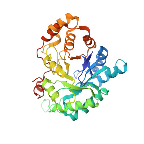
    A high-throughput screen identified 3-(3,4-dihydroisoquinolin-2(1H)-ylsulfonyl)benzoic acid as a novel, highly potent (low nM), and isoform-selective (1500-fold) inhibitor of aldo-keto reductase AKR1C3: a target of interest in both breast and prostate cancer. Crystal structure studies showed that the carboxylate group occupies the oxyanion hole in the enzyme, while the sulfonamide provides the correct twist to allow the dihydroisoquinoline to bind in an adjacent hydrophobic pocket. SAR studies around this lead showed that the positioning of the carboxylate was critical, although it could be substituted by acid isosteres and amides. Small substituents on the dihydroisoquinoline gave improvements in potency. A set of "reverse sulfonamides" showed a 12-fold preference for the R stereoisomer. The compounds showed good cellular potency, as measured by inhibition of AKR1C3 metabolism of a known dinitrobenzamide substrate, with a broad rank order between enzymic and cellular activity, but amide analogues were more effective than predicted by the cellular assay.


  • Organizational Affiliation

    Auckland Cancer Society Research Centre, The University of Auckland, Private Bag 92019, Auckland 1142, New Zealand.


Macromolecules
Find similar proteins by:  (by identity cutoff)  |  3D Structure
Entity ID: 1
MoleculeChains Sequence LengthOrganismDetailsImage
Aldo-keto reductase family 1 member C3
A, B
331Homo sapiensMutation(s): 0 
Gene Names: AKR1C3DDH1HSD17B5KIAA0119PGFS
EC: 1 (PDB Primary Data), 1.1.1.213 (PDB Primary Data), 1.1.1.112 (PDB Primary Data), 1.1.1.188 (PDB Primary Data), 1.1.1.63 (PDB Primary Data), 1.1.1.64 (PDB Primary Data), 1.3.1.20 (PDB Primary Data)
UniProt & NIH Common Fund Data Resources
Find proteins for P42330 (Homo sapiens)
Explore P42330 
Go to UniProtKB:  P42330
PHAROS:  P42330
GTEx:  ENSG00000196139 
Entity Groups  
Sequence Clusters30% Identity50% Identity70% Identity90% Identity95% Identity100% Identity
UniProt GroupP42330
Sequence Annotations
Expand
  • Reference Sequence
Small Molecules
Ligands 3 Unique
IDChains Name / Formula / InChI Key2D Diagram3D Interactions
NAP
Query on NAP

Download Ideal Coordinates CCD File 
C [auth A],
K [auth B]
NADP NICOTINAMIDE-ADENINE-DINUCLEOTIDE PHOSPHATE
C21 H28 N7 O17 P3
XJLXINKUBYWONI-NNYOXOHSSA-N
0SZ
Query on 0SZ

Download Ideal Coordinates CCD File 
D [auth A],
L [auth B]
3-(3,4-dihydroisoquinolin-2(1H)-ylsulfonyl)benzoic acid
C16 H15 N O4 S
ZGVIUMKHTXKKOX-UHFFFAOYSA-N
EDO
Query on EDO

Download Ideal Coordinates CCD File 
E [auth A]
F [auth A]
G [auth A]
H [auth A]
I [auth A]
E [auth A],
F [auth A],
G [auth A],
H [auth A],
I [auth A],
J [auth A],
M [auth B],
N [auth B],
O [auth B]
1,2-ETHANEDIOL
C2 H6 O2
LYCAIKOWRPUZTN-UHFFFAOYSA-N
Binding Affinity Annotations 
IDSourceBinding Affinity
0SZ BindingDB:  4FAM IC50: min: 13, max: 3.00e+4 (nM) from 4 assay(s)
Binding MOAD:  4FAM IC50: 13 (nM) from 1 assay(s)
Experimental Data & Validation

Experimental Data

  • Method: X-RAY DIFFRACTION
  • Resolution: 2.00 Å
  • R-Value Free: 0.218 
  • R-Value Work: 0.168 
  • R-Value Observed: 0.170 
  • Space Group: P 1 21 1
Unit Cell:
Length ( Å )Angle ( ˚ )
a = 48.717α = 90
b = 106.764β = 103.11
c = 74.882γ = 90
Software Package:
Software NamePurpose
StructureStudiodata collection
PHASERphasing
REFMACrefinement

Structure Validation

View Full Validation Report


Ligand Structure Quality Assessment 


Entry History 

Revision History  (Full details and data files)

  • Version 1.0: 2012-10-10
    Type: Initial release
  • Version 1.1: 2023-09-13
    Changes: Data collection, Database references, Derived calculations, Refinement description