4EMO

Crystal structure of the PH domain of SHARPIN


Experimental Data Snapshot

  • Method: X-RAY DIFFRACTION
  • Resolution: 2.00 Å
  • R-Value Free: 0.269 
  • R-Value Work: 0.209 
  • R-Value Observed: 0.212 

wwPDB Validation   3D Report Full Report


This is version 1.2 of the entry. See complete history


Literature

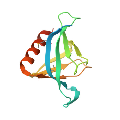
Structural Analysis of SHARPIN, a Subunit of a Large Multi-protein E3 Ubiquitin Ligase, Reveals a Novel Dimerization Function for the Pleckstrin Homology Superfold.

Stieglitz, B.Haire, L.F.Dikic, I.Rittinger, K.

(2012) J Biol Chem 287: 20823-20829

  • DOI: https://doi.org/10.1074/jbc.M112.359547
  • Primary Citation of Related Structures:  
    4EMO

  • PubMed Abstract: 

    SHARPIN (SHANK-associated RH domain interacting protein) is part of a large multi-protein E3 ubiquitin ligase complex called LUBAC (linear ubiquitin chain assembly complex), which catalyzes the formation of linear ubiquitin chains and regulates immune and apoptopic signaling pathways. The C-terminal half of SHARPIN contains ubiquitin-like domain and Npl4-zinc finger domains that mediate the interaction with the LUBAC subunit HOIP and ubiquitin, respectively. In contrast, the N-terminal region does not show any homology with known protein interaction domains but has been suggested to be responsible for self-association of SHARPIN, presumably via a coiled-coil region. We have determined the crystal structure of the N-terminal portion of SHARPIN, which adopts the highly conserved pleckstrin homology superfold that is often used as a scaffold to create protein interaction modules. We show that in SHARPIN, this domain does not appear to be used as a ligand recognition domain because it lacks many of the surface properties that are present in other pleckstrin homology fold-based interaction modules. Instead, it acts as a dimerization module extending the functional applications of this superfold.


  • Organizational Affiliation

    Division of Molecular Structure, Medical Research Council National Institute for Medical Research, The Ridgeway, London NW7 1AA, United Kingdom.


Macromolecules
Find similar proteins by:  (by identity cutoff)  |  3D Structure
Entity ID: 1
MoleculeChains Sequence LengthOrganismDetailsImage
Sharpin
A, B, C, D
129Homo sapiensMutation(s): 2 
Gene Names: PSEC0216SHARPINSIPL1
UniProt & NIH Common Fund Data Resources
Find proteins for Q9H0F6 (Homo sapiens)
Explore Q9H0F6 
Go to UniProtKB:  Q9H0F6
PHAROS:  Q9H0F6
GTEx:  ENSG00000179526 
Entity Groups  
Sequence Clusters30% Identity50% Identity70% Identity90% Identity95% Identity100% Identity
UniProt GroupQ9H0F6
Sequence Annotations
Expand
  • Reference Sequence
Small Molecules
Modified Residues  1 Unique
IDChains TypeFormula2D DiagramParent
MSE
Query on MSE
A, B, C, D
L-PEPTIDE LINKINGC5 H11 N O2 SeMET
Experimental Data & Validation

Experimental Data

  • Method: X-RAY DIFFRACTION
  • Resolution: 2.00 Å
  • R-Value Free: 0.269 
  • R-Value Work: 0.209 
  • R-Value Observed: 0.212 
  • Space Group: P 43 21 2
Unit Cell:
Length ( Å )Angle ( ˚ )
a = 61.55α = 90
b = 61.55β = 90
c = 222.813γ = 90
Software Package:
Software NamePurpose
DENZOdata reduction
SCALEPACKdata scaling
RESOLVEphasing
REFMACrefinement
PDB_EXTRACTdata extraction
ADSCdata collection
PHENIXphasing

Structure Validation

View Full Validation Report


Entry History 

Deposition Data

Revision History  (Full details and data files)

  • Version 1.0: 2012-05-02
    Type: Initial release
  • Version 1.1: 2012-05-16
    Changes: Database references
  • Version 1.2: 2012-07-25
    Changes: Database references