Structural and biochemical characterization of human mitochondrial branched-chain alpha-ketoacid dehydrogenase phosphatase.
Wynn, R.M., Li, J., Brautigam, C.A., Chuang, J.L., Chuang, D.T.(2012) J Biol Chem 287: 9178-9192
- PubMed: 22291014
- DOI: https://doi.org/10.1074/jbc.M111.314963
- Primary Citation of Related Structures:
4DA1 - PubMed Abstract:
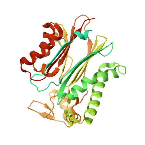
The branched-chain α-ketoacid dehydrogenase phosphatase (BDP) component of the human branched-chain α-ketoacid dehydrogenase complex (BCKDC) has been expressed in Escherichia coli and purified in the soluble form. The monomeric BDP shows a strict dependence on Mn(2+) ions for phosphatase activity, whereas Mg(2+) and Ca(2+) ions do not support catalysis. Metal binding constants for BDP, determined by competition isothermal titration calorimetry, are 2.4 nm and 10 μm for Mn(2+) and Mg(2+) ions, respectively. Using the phosphorylated decarboxylase component (p-E1b) of BCKDC as a substrate, BDP shows a specific activity of 68 nmol/min/mg. The Ca(2+)-independent binding of BDP to the 24-meric transacylase (dihydrolipoyl transacylase; E2b) core of BCKDC results in a 3-fold increase in the dephosphorylation rate of p-E1b. However, the lipoyl prosthetic group on E2b is not essential for BDP binding or E2b-stimulated phosphatase activity. Acidic residues in the C-terminal linker of the E2b lipoyl domain are essential for the interaction between BDP and E2b. The BDP structure was determined by x-ray crystallography to 2.4 Å resolution. The BDP structure is dominated by a central β-sandwich. There are two protrusions forming a narrow cleft ∼10 Å wide, which constitutes the active site. The carboxylate moieties of acidic residues Asp-109, Asp-207, Asp-298, and Asp-337 in the active-site cleft participate in binding two metal ions. Substitutions of these residues with alanine nullify BDP phosphatase activity. Alteration of the nearby Arg-104 increases the K(m) for p-E1b peptide by 60-fold, suggesting that this residue is critical for the recognition of the native p-E1b protein.
Organizational Affiliation:
Department of Biochemistry, University of Texas Southwestern Medical Center at Dallas, Dallas, Texas 75390-9038, USA. richard.wynn@utsouthwestern.edu