4BTB

CRYSTAL STRUCTURE OF THE PEPTIDE(PRO)9 BOUND COMPLEX OF N-TERMINAL DOMAIN AND PEPTIDE SUBSTRATE BINDING DOMAIN OF PROLYL-4 HYDROXYLASE (RESIDUES 1-238) TYPE I FROM HUMAN


Experimental Data Snapshot

  • Method: X-RAY DIFFRACTION
  • Resolution: 1.90 Å
  • R-Value Free: 0.235 
  • R-Value Work: 0.183 
  • R-Value Observed: 0.185 

wwPDB Validation   3D Report Full Report


This is version 1.5 of the entry. See complete history


Literature

The Structural Motifs for Substrate Binding and Dimerization of the Alpha Subunit of Collagen Prolyl 4-Hydroxylase

Anantharajan, J.Koski, M.K.Kursula, P.Hieta, R.Bergmann, U.Myllyharju, J.Wierenga, R.K.

(2013) Structure 21: 2107

  • DOI: https://doi.org/10.1016/j.str.2013.09.005
  • Primary Citation of Related Structures:  
    2YQ8, 4BT8, 4BT9, 4BTA, 4BTB

  • PubMed Abstract: 

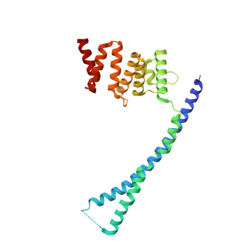
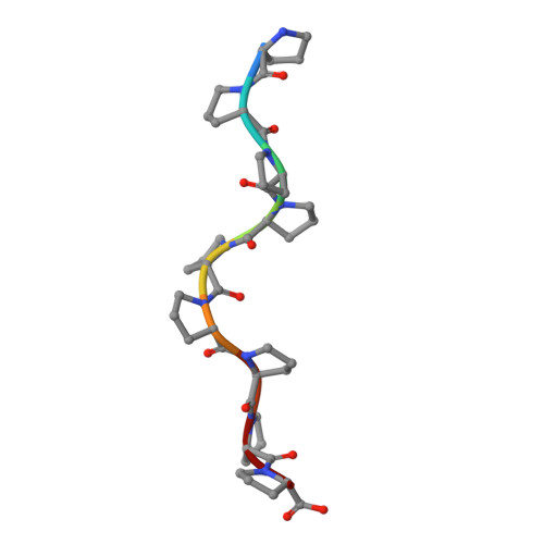
    Collagen prolyl 4-hydroxylase (C-P4H) catalyzes the proline hydroxylation of procollagen, an essential modification in the maturation of collagens. C-P4H consists of two catalytic α subunits and two protein disulfide isomerase β subunits. The assembly of these subunits is unknown. The α subunit contains an N domain (1-143), a peptide-substrate-binding-domain (PSB, 144-244) and a catalytic domain (245-517). Here, we report the dimeric structure of the N-terminal region (1-244) of the α subunit. It is shown that the N domain has an important role in the assembly of the C-P4H tetramer, by forming an extended four-helix bundle that includes an antiparallel coiled-coil dimerization motif between the two α subunits. Complexes of this construct with a C-P4H inhibitor and substrate show the mode of peptide-binding to the PSB domain. Both peptides adopt a poly-(L)-proline-type-II helix conformation and bind in a curved, asymmetric groove lined by conserved tyrosines and an Arg-Asp salt bridge.


  • Organizational Affiliation

    Biocenter Oulu and Department of Biochemistry, University of Oulu, P.O. Box 3000, FIN-90014 Oulu, Finland.


Macromolecules
Find similar proteins by:  (by identity cutoff)  |  3D Structure
Entity ID: 1
MoleculeChains Sequence LengthOrganismDetailsImage
PROLYL 4-HYDROXYLASE SUBUNIT ALPHA-1239Homo sapiensMutation(s): 0 
EC: 1.14.11.2
UniProt & NIH Common Fund Data Resources
Find proteins for P13674 (Homo sapiens)
Explore P13674 
Go to UniProtKB:  P13674
PHAROS:  P13674
GTEx:  ENSG00000122884 
Entity Groups  
Sequence Clusters30% Identity50% Identity70% Identity90% Identity95% Identity100% Identity
UniProt GroupP13674
Sequence Annotations
Expand
  • Reference Sequence

Find similar proteins by:  Sequence   |   3D Structure  

Entity ID: 2
MoleculeChains Sequence LengthOrganismDetailsImage
POLY PROLINE PEPTIDEB [auth C]9synthetic constructMutation(s): 0 
Sequence Annotations
Expand
  • Reference Sequence
Experimental Data & Validation

Experimental Data

  • Method: X-RAY DIFFRACTION
  • Resolution: 1.90 Å
  • R-Value Free: 0.235 
  • R-Value Work: 0.183 
  • R-Value Observed: 0.185 
  • Space Group: C 1 2 1
Unit Cell:
Length ( Å )Angle ( ˚ )
a = 76.93α = 90
b = 53.043β = 95.84
c = 79.928γ = 90
Software Package:
Software NamePurpose
PHENIXrefinement
SAINTdata reduction
SADABSdata scaling
PHASERphasing

Structure Validation

View Full Validation Report


Entry History 

Deposition Data

Revision History  (Full details and data files)

  • Version 1.0: 2013-10-09
    Type: Initial release
  • Version 1.1: 2013-10-16
    Changes: Database references
  • Version 1.2: 2013-11-20
    Changes: Database references
  • Version 1.3: 2013-12-25
    Changes: Database references
  • Version 1.4: 2017-07-05
    Changes: Data collection
  • Version 1.5: 2023-12-20
    Changes: Data collection, Database references, Other, Refinement description