4AZA

Improved eIF4E binding peptides by phage display guided design.


Experimental Data Snapshot

  • Method: X-RAY DIFFRACTION
  • Resolution: 2.16 Å
  • R-Value Free: 0.279 
  • R-Value Work: 0.233 
  • R-Value Observed: 0.235 

wwPDB Validation   3D Report Full Report


Ligand Structure Quality Assessment 


This is version 1.2 of the entry. See complete history


Literature

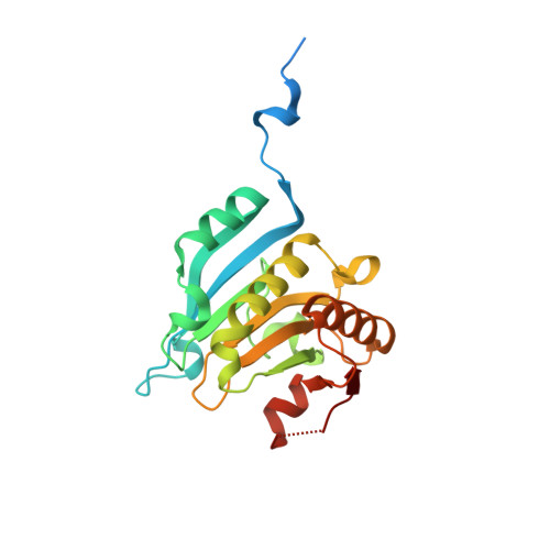
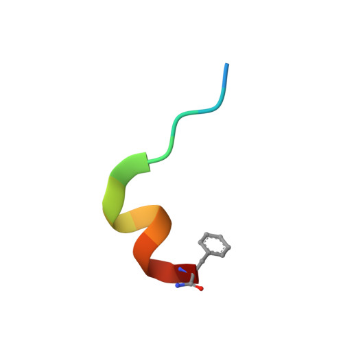
Improved Eif4E Binding Peptides by Phage Display Guided Design: Plasticity of Interacting Surfaces Yield Collective Effects.

Zhou, W.Quah, S.T.Verma, C.S.Liu, Y.Lane, D.P.Brown, C.J.

(2012) PLoS One 7: 47235

  • DOI: https://doi.org/10.1371/journal.pone.0047235
  • Primary Citation of Related Structures:  
    4AZA

  • PubMed Abstract: 

    Eukaryotic initiation factor (eIF)4E is over-expressed in many types of cancer such as breast, head and neck, and lung. A consequence of increased levels of eIF4E is the preferential translation of pro-tumorigenic proteins (e.g. c-Myc and vascular endothelial growth factor) and as a result is regarded as a potential therapeutic target. In this work a novel phage display peptide has been isolated against eIF4E. From the phage sequence two amino acids were delineated which improved binding when substituted into the eIF4G1 sequence. Neither of these substitutions were involved in direct interactions with eIF4E and acted either via optimization of the helical capping motif or restricting the conformational flexibility of the peptide. In contrast, substitutions of the remaining phage derived amino acids into the eIF4G1 sequence disrupted binding of the peptide to eIF4E. Interestingly when some of these disruptive substitutions were combined with key mutations from the phage peptide, they lead to improved affinities. Atomistic computer simulations revealed that the phage and the eIF4G1 derivative peptide sequences differ subtly in their interaction sites on eIF4E. This raises the issue, especially in the context of planar interaction sites such as those exhibited by eIF4E, that given the intricate plasticity of protein surfaces, the construction of structure-activity relationships should account for the possibility of significant movement in the spatial positioning of the peptide binding interface, including significant librational motions of the peptide.


  • Organizational Affiliation

    Bioinformatics Institute, Agency for Science, Technology and Research (ASTAR), Singapore.


Macromolecules
Find similar proteins by:  (by identity cutoff)  |  3D Structure
Entity ID: 1
MoleculeChains Sequence LengthOrganismDetailsImage
EUKARYOTIC TRANSLATION INITIATION FACTOR 4E
A, C
217Homo sapiensMutation(s): 0 
UniProt & NIH Common Fund Data Resources
Find proteins for P06730 (Homo sapiens)
Explore P06730 
Go to UniProtKB:  P06730
PHAROS:  P06730
GTEx:  ENSG00000151247 
Entity Groups  
Sequence Clusters30% Identity50% Identity70% Identity90% Identity95% Identity100% Identity
UniProt GroupP06730
Sequence Annotations
Expand
  • Reference Sequence

Find similar proteins by:  Sequence   |   3D Structure  

Entity ID: 2
MoleculeChains Sequence LengthOrganismDetailsImage
EIF4G1_D5S PEPTIDE
B, D
14Homo sapiensMutation(s): 0 
UniProt & NIH Common Fund Data Resources
Find proteins for Q04637 (Homo sapiens)
Explore Q04637 
Go to UniProtKB:  Q04637
PHAROS:  Q04637
GTEx:  ENSG00000114867 
Entity Groups  
Sequence Clusters30% Identity50% Identity70% Identity90% Identity95% Identity100% Identity
UniProt GroupQ04637
Sequence Annotations
Expand
  • Reference Sequence
Small Molecules
Ligands 1 Unique
IDChains Name / Formula / InChI Key2D Diagram3D Interactions
MGO
Query on MGO

Download Ideal Coordinates CCD File 
E [auth A],
F [auth C]
[[(2R,3S,4R,5R)-5-(6-AMINO-3-METHYL-4-OXO-5H-IMIDAZO[4,5-C]PYRIDIN-1-YL)-3,4-DIHYDROXY-OXOLAN-2-YL]METHOXY-HYDROXY-PHOSPHORYL] PHOSPHONO HYDROGEN PHOSPHATE
C12 H20 N4 O14 P3
LPHBJZOLBGBNJF-XYHAGOFUSA-O
Experimental Data & Validation

Experimental Data

  • Method: X-RAY DIFFRACTION
  • Resolution: 2.16 Å
  • R-Value Free: 0.279 
  • R-Value Work: 0.233 
  • R-Value Observed: 0.235 
  • Space Group: P 1 21 1
Unit Cell:
Length ( Å )Angle ( ˚ )
a = 60.662α = 90
b = 38.168β = 90
c = 121.758γ = 90
Software Package:
Software NamePurpose
REFMACrefinement
PROTEUM2data reduction
PROTEUM2data scaling
PHASERphasing

Structure Validation

View Full Validation Report


Ligand Structure Quality Assessment 


Entry History 

Deposition Data

Revision History  (Full details and data files)

  • Version 1.0: 2012-08-08
    Type: Initial release
  • Version 1.1: 2013-03-06
    Changes: Database references
  • Version 1.2: 2023-12-20
    Changes: Data collection, Database references, Derived calculations, Other, Refinement description