3ZZY

Crystal structure of a Raver1 PRI3 peptide in complex with polypyrimidine tract binding protein RRM2


Experimental Data Snapshot

  • Method: X-RAY DIFFRACTION
  • Resolution: 1.40 Å
  • R-Value Free: 0.234 
  • R-Value Work: 0.224 
  • R-Value Observed: 0.224 

wwPDB Validation   3D Report Full Report


This is version 1.2 of the entry. See complete history


Literature

Crystallographic Analysis of Polypyrimidine Tract-Binding Protein-Raver1 Interactions Involved in Regulation of Alternative Splicing.

Joshi, A.Coelho, M.B.Kotik-Kogan, O.Simpson, P.J.Matthews, S.J.Smith, C.W.Curry, S.

(2011) Structure 19: 1816

  • DOI: https://doi.org/10.1016/j.str.2011.09.020
  • Primary Citation of Related Structures:  
    3ZZY, 3ZZZ

  • PubMed Abstract: 

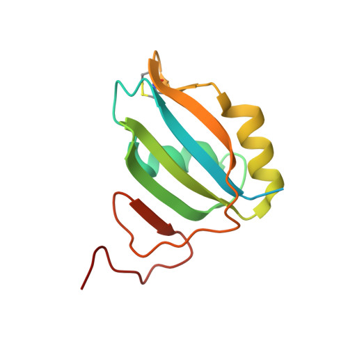
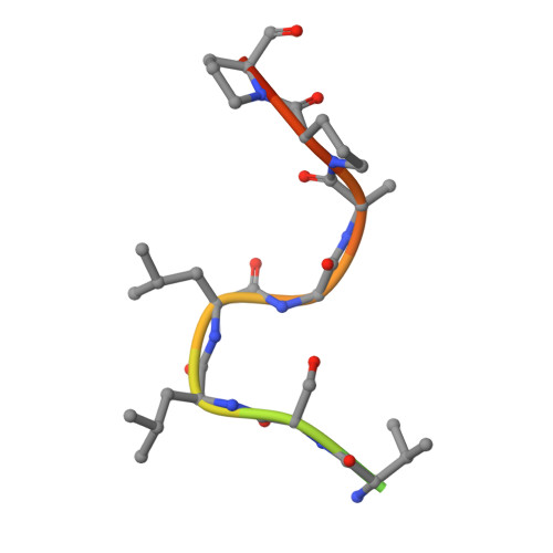
    The polypyrimidine tract-binding protein (PTB) is an important regulator of alternative splicing. PTB-regulated splicing of α-tropomyosin is enhanced by Raver1, a protein with four PTB-Raver1 interacting motifs (PRIs) that bind to the helical face of the second RNA recognition motif (RRM2) in PTB. We present the crystal structures of RRM2 in complex with PRI3 and PRI4 from Raver1, which--along with structure-based mutagenesis--reveal the molecular basis of their differential binding. High-affinity binding by Raver1 PRI3 involves shape-matched apolar contacts complemented by specific hydrogen bonds, a new variant of an established mode of peptide-RRM interaction. Our results refine the sequence of the PRI motif and place important structural constraints on functional models of PTB-Raver1 interactions. Our analysis indicates that the observed Raver1-PTB interaction is a general mode of binding that applies to Raver1 complexes with PTB paralogues such as nPTB and to complexes of Raver2 with PTB.


  • Organizational Affiliation

    Division of Cell and Molecular Biology, Imperial College, Exhibition Road, London SW7 2AZ, UK.


Macromolecules
Find similar proteins by:  (by identity cutoff)  |  3D Structure
Entity ID: 1
MoleculeChains Sequence LengthOrganismDetailsImage
POLYPYRIMIDINE TRACT-BINDING PROTEIN 1
A, B
130Homo sapiensMutation(s): 0 
UniProt & NIH Common Fund Data Resources
Find proteins for P26599 (Homo sapiens)
Explore P26599 
Go to UniProtKB:  P26599
PHAROS:  P26599
GTEx:  ENSG00000011304 
Entity Groups  
Sequence Clusters30% Identity50% Identity70% Identity90% Identity95% Identity100% Identity
UniProt GroupP26599
Sequence Annotations
Expand
  • Reference Sequence

Find similar proteins by:  Sequence   |   3D Structure  

Entity ID: 2
MoleculeChains Sequence LengthOrganismDetailsImage
RIBONUCLEOPROTEIN PTB-BINDING 1
C, D
16Mus musculusMutation(s): 1 
UniProt & NIH Common Fund Data Resources
Find proteins for Q9CW46 (Mus musculus)
Explore Q9CW46 
Go to UniProtKB:  Q9CW46
IMPC:  MGI:1919016
Entity Groups  
Sequence Clusters30% Identity50% Identity70% Identity90% Identity95% Identity100% Identity
UniProt GroupQ9CW46
Sequence Annotations
Expand
  • Reference Sequence
Experimental Data & Validation

Experimental Data

  • Method: X-RAY DIFFRACTION
  • Resolution: 1.40 Å
  • R-Value Free: 0.234 
  • R-Value Work: 0.224 
  • R-Value Observed: 0.224 
  • Space Group: C 1 2 1
Unit Cell:
Length ( Å )Angle ( ˚ )
a = 74.233α = 90
b = 60.606β = 107.51
c = 60.842γ = 90
Software Package:
Software NamePurpose
CNSrefinement
MOSFLMdata reduction
SCALAdata scaling
PHASERphasing

Structure Validation

View Full Validation Report


Entry History 

Deposition Data

Revision History  (Full details and data files)

  • Version 1.0: 2011-12-28
    Type: Initial release
  • Version 1.1: 2019-07-17
    Changes: Data collection
  • Version 1.2: 2023-12-20
    Changes: Data collection, Database references, Other, Refinement description