3ZQS

Human FANCL central domain


Experimental Data Snapshot

  • Method: X-RAY DIFFRACTION
  • Resolution: 2.00 Å
  • R-Value Free: 0.207 
  • R-Value Work: 0.183 
  • R-Value Observed: 0.184 

wwPDB Validation   3D Report Full Report


This is version 1.3 of the entry. See complete history


Literature

Structural Analysis of Human Fancl, the E3 Ligase in the Fanconi Anemia Pathway.

Hodson, C.Cole, A.R.Lewis, L.P.Miles, J.A.Purkiss-Trew, A.Walden, H.

(2011) J Biol Chem 286: 32628

  • DOI: https://doi.org/10.1074/jbc.M111.244632
  • Primary Citation of Related Structures:  
    3ZQS

  • PubMed Abstract: 

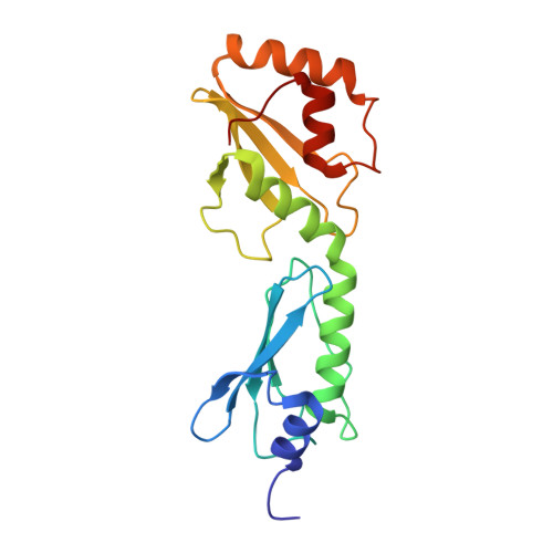
    The Fanconi anemia (FA) pathway is essential for the repair of DNA interstrand cross-links. At the heart of this pathway is the monoubiquitination of the FANCI-FANCD2 (ID) complex by the multiprotein "core complex" containing the E3 ubiquitin ligase FANCL. Vertebrate organisms have the eight-protein core complex, whereas invertebrates apparently do not. We report here the structure of the central domain of human FANCL in comparison with the recently solved Drosophila melanogaster FANCL. Our data represent the first structural detail into the catalytic core of the human system and reveal that the central fold of FANCL is conserved between species. However, there are macromolecular differences between the FANCL proteins that may account for the apparent distinctions in core complex requirements between the vertebrate and invertebrate FA pathways. In addition, we characterize the binding of human FANCL with its partners, Ube2t, FANCD2, and FANCI. Mutational analysis reveals which residues are required for substrate binding, and we also show the domain required for E2 binding.


  • Organizational Affiliation

    Protein Structure and Function Laboratory, Lincoln's Inn Fields Laboratories of the London Research Institute, Cancer Research UK, 44 Lincoln's Inn Fields, London WC2A 3LY, United Kingdom.


Macromolecules
Find similar proteins by:  (by identity cutoff)  |  3D Structure
Entity ID: 1
MoleculeChains Sequence LengthOrganismDetailsImage
E3 UBIQUITIN-PROTEIN LIGASE FANCL
A, B
186Homo sapiensMutation(s): 0 
EC: 6.3.2.19
UniProt & NIH Common Fund Data Resources
Find proteins for Q9NW38 (Homo sapiens)
Explore Q9NW38 
Go to UniProtKB:  Q9NW38
PHAROS:  Q9NW38
GTEx:  ENSG00000115392 
Entity Groups  
Sequence Clusters30% Identity50% Identity70% Identity90% Identity95% Identity100% Identity
UniProt GroupQ9NW38
Sequence Annotations
Expand
  • Reference Sequence
Small Molecules
Ligands 3 Unique
IDChains Name / Formula / InChI Key2D Diagram3D Interactions
P6G
Query on P6G

Download Ideal Coordinates CCD File 
D [auth A]
E [auth A]
F [auth A]
G [auth A]
H [auth A]
D [auth A],
E [auth A],
F [auth A],
G [auth A],
H [auth A],
I [auth A],
J [auth A],
N [auth B],
O [auth B],
P [auth B]
HEXAETHYLENE GLYCOL
C12 H26 O7
IIRDTKBZINWQAW-UHFFFAOYSA-N
PRO
Query on PRO

Download Ideal Coordinates CCD File 
C [auth A],
K [auth B]
PROLINE
C5 H9 N O2
ONIBWKKTOPOVIA-BYPYZUCNSA-N
NA
Query on NA

Download Ideal Coordinates CCD File 
L [auth B],
M [auth B]
SODIUM ION
Na
FKNQFGJONOIPTF-UHFFFAOYSA-N
Experimental Data & Validation

Experimental Data

  • Method: X-RAY DIFFRACTION
  • Resolution: 2.00 Å
  • R-Value Free: 0.207 
  • R-Value Work: 0.183 
  • R-Value Observed: 0.184 
  • Space Group: C 1 2 1
Unit Cell:
Length ( Å )Angle ( ˚ )
a = 147.6α = 90
b = 102.52β = 94.09
c = 65.76γ = 90
Software Package:
Software NamePurpose
PHENIXrefinement
XDSdata reduction
XSCALEdata scaling
PHASERphasing

Structure Validation

View Full Validation Report


Entry History 

Deposition Data

Revision History  (Full details and data files)

  • Version 1.0: 2011-07-20
    Type: Initial release
  • Version 1.1: 2011-08-03
    Changes: Database references, Structure summary
  • Version 1.2: 2011-09-21
    Changes: Database references
  • Version 1.3: 2023-12-20
    Changes: Data collection, Database references, Derived calculations, Other, Refinement description