3ZDL

Vinculin head (1-258) in complex with a RIAM fragment


Experimental Data Snapshot

  • Method: X-RAY DIFFRACTION
  • Resolution: 2.30 Å
  • R-Value Free: 0.230 
  • R-Value Work: 0.188 
  • R-Value Observed: 0.190 

wwPDB Validation   3D Report Full Report


This is version 1.5 of the entry. See complete history


Literature

Riam and Vinculin Binding to Talin are Mutually Exclusive and Regulate Adhesion Assembly and Turnover.

Goult, B.T.Zacharchenko, T.Bate, N.Tsang, R.Hey, F.Gingras, A.R.Elliott, P.R.Roberts, G.C.Ballestrem, C.Critchley, D.R.Barsukov, I.L.

(2013) J Biol Chem 288: 8238

  • DOI: https://doi.org/10.1074/jbc.M112.438119
  • Primary Citation of Related Structures:  
    2L10, 2L7A, 2L7N, 2LQG, 3ZDL

  • PubMed Abstract: 

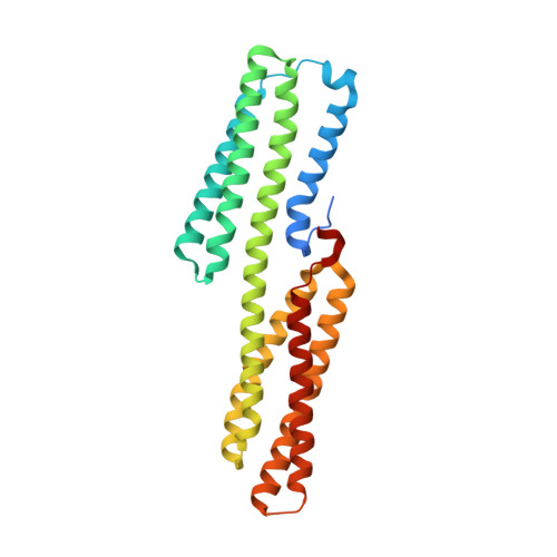
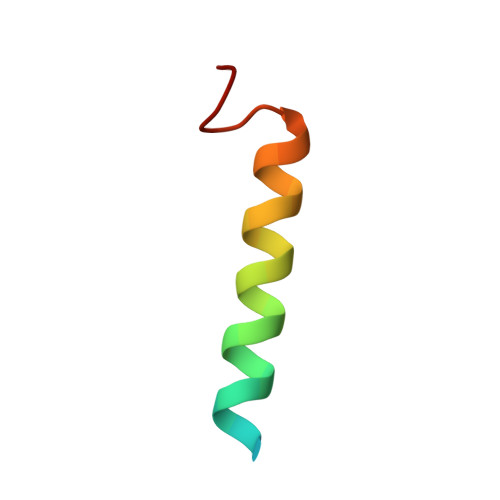
    Talin activates integrins, couples them to F-actin, and recruits vinculin to focal adhesions (FAs). Here, we report the structural characterization of the talin rod: 13 helical bundles (R1-R13) organized into a compact cluster of four-helix bundles (R2-R4) within a linear chain of five-helix bundles. Nine of the bundles contain vinculin-binding sites (VBS); R2R3 are atypical, with each containing two VBS. Talin R2R3 also binds synergistically to RIAM, a Rap1 effector involved in integrin activation. Biochemical and structural data show that vinculin and RIAM binding to R2R3 is mutually exclusive. Moreover, vinculin binding requires domain unfolding, whereas RIAM binds the folded R2R3 double domain. In cells, RIAM is enriched in nascent adhesions at the leading edge whereas vinculin is enriched in FAs. We propose a model in which RIAM binding to R2R3 initially recruits talin to membranes where it activates integrins. As talin engages F-actin, force exerted on R2R3 disrupts RIAM binding and exposes the VBS, which recruit vinculin to stabilize the complex.


  • Organizational Affiliation

    Department of Biochemistry, University of Leicester, Lancaster Road, Leicester LE1 9HN, United Kingdom.


Macromolecules
Find similar proteins by:  (by identity cutoff)  |  3D Structure
Entity ID: 1
MoleculeChains Sequence LengthOrganismDetailsImage
VINCULIN280Gallus gallusMutation(s): 0 
UniProt
Find proteins for P12003 (Gallus gallus)
Explore P12003 
Go to UniProtKB:  P12003
Entity Groups  
Sequence Clusters30% Identity50% Identity70% Identity90% Identity95% Identity100% Identity
UniProt GroupP12003
Sequence Annotations
Expand
  • Reference Sequence
Find similar proteins by:  (by identity cutoff)  |  3D Structure
Entity ID: 2
MoleculeChains Sequence LengthOrganismDetailsImage
AMYLOID BETA A4 PRECURSOR PROTEIN-BINDING FAMILY B MEMBER 1-INTERACTING PROTEIN32Homo sapiensMutation(s): 1 
UniProt & NIH Common Fund Data Resources
Find proteins for Q7Z5R6 (Homo sapiens)
Explore Q7Z5R6 
Go to UniProtKB:  Q7Z5R6
PHAROS:  Q7Z5R6
GTEx:  ENSG00000077420 
Entity Groups  
Sequence Clusters30% Identity50% Identity70% Identity90% Identity95% Identity100% Identity
UniProt GroupQ7Z5R6
Sequence Annotations
Expand
  • Reference Sequence
Experimental Data & Validation

Experimental Data

  • Method: X-RAY DIFFRACTION
  • Resolution: 2.30 Å
  • R-Value Free: 0.230 
  • R-Value Work: 0.188 
  • R-Value Observed: 0.190 
  • Space Group: P 21 21 2
Unit Cell:
Length ( Å )Angle ( ˚ )
a = 51.73α = 90
b = 70.03β = 90
c = 95.6γ = 90
Software Package:
Software NamePurpose
PHENIXrefinement
iMOSFLMdata reduction
SCALAdata scaling
PHASERphasing

Structure Validation

View Full Validation Report


Entry History 

Deposition Data

Revision History  (Full details and data files)

  • Version 1.0: 2013-02-13
    Type: Initial release
  • Version 1.1: 2013-02-20
    Changes: Database references
  • Version 1.2: 2013-03-06
    Changes: Derived calculations, Other
  • Version 1.3: 2013-04-10
    Changes: Derived calculations, Other
  • Version 1.4: 2013-04-17
    Changes: Database references
  • Version 1.5: 2023-12-20
    Changes: Data collection, Database references, Other, Refinement description