3WHT

Crystal structure of ERGIC-53/MCFD2, Calcium-free form


Experimental Data Snapshot

  • Method: X-RAY DIFFRACTION
  • Resolution: 1.80 Å
  • R-Value Free: 0.202 
  • R-Value Work: 0.175 
  • R-Value Observed: 0.177 

wwPDB Validation   3D Report Full Report


This is version 1.2 of the entry. See complete history


Literature

Structural Basis for Disparate Sugar-Binding Specificities in the Homologous Cargo Receptors ERGIC-53 and VIP36

Satoh, T.Suzuki, K.Yamaguchi, T.Kato, K.

(2014) PLoS One 9: e87963-e87963

  • DOI: https://doi.org/10.1371/journal.pone.0087963
  • Primary Citation of Related Structures:  
    3WHT, 3WHU, 3WNX

  • PubMed Abstract: 

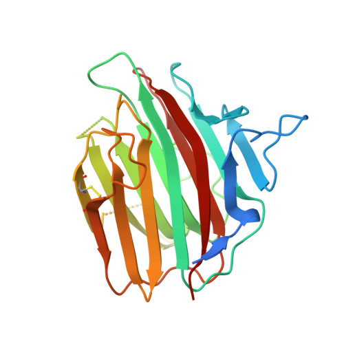
    ERGIC-53 and VIP36 are categorized as leguminous type (L-type) lectins, and they function as cargo receptors for trafficking certain N-linked glycoproteins in the secretory pathway in animal cells. They share structural similarities in their carbohydrate recognition domains (CRDs) but exhibit distinct sugar-binding specificities and affinities. VIP36 specifically interacts with the α1,2-linked D1 mannosyl arm without terminal glucosylation, while ERGIC-53 shows a broader specificity and lower binding affinity to the high-mannose-type oligosaccharides, irrespective of the presence or absence of the non-reducing terminal glucose residue at the D1 arm. In this study, we determined the crystal structure of ERGIC-53-CRD in complex with their binding partner, MCFD2 and the α1,2 mannotriose which corresponds to the trisaccharide of the D1 arm of high-mannose-type glycans. ERGIC-53 can interact with the D1 trimannosyl arm in two alternative modes, one of which is similar but distinct from that previously observed for VIP36. ERGIC-53 has a shallower sugar-binding pocket than VIP36 because of the single amino acid substitution, Asp-to-Gly. This enables ERGIC-53 to accommodate the non-reducing terminal glucose of the D1 arm in its CRD. In the other interaction mode, the 3-OH group of the terminal mannose was situated outward with respect to the sugar binding pocket, also enabling the Glcα1-3 linkage formation without steric hindrance. Our findings thus provide a structural basis for the broad sugar-binding specificity of the ERGIC-53/MCFD2 cargo receptor complex.


  • Organizational Affiliation

    Graduate School of Pharmaceutical Sciences, Nagoya City University, Mizuho-ku, Nagoya, Japan ; JST, PRESTO, Mizuho-ku, Nagoya, Japan.


Macromolecules
Find similar proteins by:  (by identity cutoff)  |  3D Structure
Entity ID: 1
MoleculeChains Sequence LengthOrganismDetailsImage
Protein ERGIC-53246Homo sapiensMutation(s): 0 
Gene Names: ERGIC53
UniProt & NIH Common Fund Data Resources
Find proteins for P49257 (Homo sapiens)
Explore P49257 
Go to UniProtKB:  P49257
PHAROS:  P49257
GTEx:  ENSG00000074695 
Entity Groups  
Sequence Clusters30% Identity50% Identity70% Identity90% Identity95% Identity100% Identity
UniProt GroupP49257
Sequence Annotations
Expand
  • Reference Sequence
Find similar proteins by:  (by identity cutoff)  |  3D Structure
Entity ID: 2
MoleculeChains Sequence LengthOrganismDetailsImage
Multiple coagulation factor deficiency protein 2104Homo sapiensMutation(s): 0 
Gene Names: MCFD2
UniProt & NIH Common Fund Data Resources
Find proteins for Q8NI22 (Homo sapiens)
Explore Q8NI22 
Go to UniProtKB:  Q8NI22
PHAROS:  Q8NI22
GTEx:  ENSG00000180398 
Entity Groups  
Sequence Clusters30% Identity50% Identity70% Identity90% Identity95% Identity100% Identity
UniProt GroupQ8NI22
Sequence Annotations
Expand
  • Reference Sequence
Experimental Data & Validation

Experimental Data

  • Method: X-RAY DIFFRACTION
  • Resolution: 1.80 Å
  • R-Value Free: 0.202 
  • R-Value Work: 0.175 
  • R-Value Observed: 0.177 
  • Space Group: C 1 2 1
Unit Cell:
Length ( Å )Angle ( ˚ )
a = 103.99α = 90
b = 59.76β = 104.9
c = 55.07γ = 90
Software Package:
Software NamePurpose
HKL-2000data collection
MOLREPphasing
REFMACrefinement
DENZOdata reduction
SCALEPACKdata scaling

Structure Validation

View Full Validation Report


Entry History 

Deposition Data

Revision History  (Full details and data files)

  • Version 1.0: 2014-01-15
    Type: Initial release
  • Version 1.1: 2014-02-19
    Changes: Database references
  • Version 1.2: 2023-11-08
    Changes: Data collection, Database references, Derived calculations, Refinement description