


3VJL
Crystal structure of human depiptidyl peptidase IV (DPP-4) in complex with a prolylthiazolidine inhibitor #2
- PDB DOI: https://doi.org/10.2210/pdb3VJL/pdb
- Classification: HYDROLASE/HYDROLASE INHIBITOR
- Organism(s): Homo sapiens
- Expression System: Spodoptera frugiperda
- Mutation(s): No
- Deposited: 2011-10-24 Released: 2012-10-24
Experimental Data Snapshot
- Method: X-RAY DIFFRACTION
- Resolution: 2.39 Å
- R-Value Free: 0.230
- R-Value Work: 0.184
- R-Value Observed: 0.186

This is version 2.1 of the entry. See complete history.

Macromolecules
Find similar proteins by:
(by identity cutoff) | 3D Structure
Entity ID: 1 | |||||
|---|---|---|---|---|---|
| Molecule | Chains | Sequence Length | Organism | Details | Image |
| Dipeptidyl peptidase 4 | 740 | Homo sapiens | Mutation(s): 0 Gene Names: DPP4, ADCP2, CD26 EC: 3.4.14.5 | 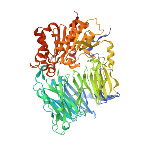 | |
UniProt & NIH Common Fund Data Resources | |||||
Find proteins for P27487 (Homo sapiens) Explore P27487 Go to UniProtKB: P27487 | |||||
PHAROS: P27487 GTEx: ENSG00000197635 | |||||
Entity Groups | |||||
| Sequence Clusters | 30% Identity50% Identity70% Identity90% Identity95% Identity100% Identity | ||||
| UniProt Group | P27487 | ||||
Sequence AnnotationsExpand | |||||
| |||||
Oligosaccharides

Small Molecules
| Ligands 2 Unique | |||||
|---|---|---|---|---|---|
| ID | Chains | Name / Formula / InChI Key | 2D Diagram | 3D Interactions | |
| W94 Query on W94 | H [auth A], L [auth B] | [(2S,4S)-4-{4-[1-phenyl-3-(trifluoromethyl)-1H-pyrazol-5-yl]piperidin-1-yl}pyrrolidin-2-yl](1,3-thiazolidin-3-yl)methanone C23 H28 F3 N5 O S CYNIHEXFNGMKLP-OALUTQOASA-N |  | ||
| NAG Query on NAG | I [auth A] J [auth A] K [auth A] M [auth B] N [auth B] | 2-acetamido-2-deoxy-beta-D-glucopyranose C8 H15 N O6 OVRNDRQMDRJTHS-FMDGEEDCSA-N |  | ||
Experimental Data & Validation
Experimental Data
- Method: X-RAY DIFFRACTION
- Resolution: 2.39 Å
- R-Value Free: 0.230
- R-Value Work: 0.184
- R-Value Observed: 0.186
- Space Group: P 21 21 21
Unit Cell:
| Length ( Å ) | Angle ( ˚ ) |
|---|---|
| a = 117.81 | α = 90 |
| b = 125.83 | β = 90 |
| c = 137.129 | γ = 90 |
| Software Name | Purpose |
|---|---|
| ADSC | data collection |
| PHASER | phasing |
| REFMAC | refinement |
| HKL-2000 | data reduction |
| SCALEPACK | data scaling |
Entry History
Deposition Data
- Released Date: 2012-10-24 Deposition Author(s): Akahoshi, F., Kishida, H., Miyaguchi, I., Yoshida, T., Ishii, S.
Revision History (Full details and data files)
- Version 1.0: 2012-10-24
Type: Initial release - Version 2.0: 2020-07-29
Type: Remediation
Reason: Carbohydrate remediation
Changes: Atomic model, Data collection, Database references, Derived calculations, Structure summary - Version 2.1: 2023-11-08
Changes: Data collection, Database references, Refinement description, Structure summary
