3V34

Crystal structure of MCPIP1 conserved domain with magnesium ion in the catalytic center


Experimental Data Snapshot

  • Method: X-RAY DIFFRACTION
  • Resolution: 2.00 Å
  • R-Value Free: 0.240 
  • R-Value Work: 0.198 
  • R-Value Observed: 0.200 

wwPDB Validation   3D Report Full Report


This is version 1.2 of the entry. See complete history


Literature

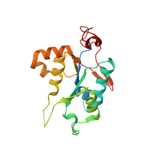
Structural study of MCPIP1 N-terminal conserved domain reveals a PIN-like RNase

Xu, J.Peng, W.Sun, Y.Wang, X.Xu, Y.Li, X.Gao, G.Rao, Z.

(2012) Nucleic Acids Res 40: 6957-6965

  • DOI: https://doi.org/10.1093/nar/gks359
  • Primary Citation of Related Structures:  
    3V32, 3V33, 3V34

  • PubMed Abstract: 

    MCP-1-induced protein 1 (MCPIP1) plays an important role in the downregulation of the LPS-induced immune response by acting as an RNase targeting IL-6 and IL-12b mRNAs. A conserved domain located in the N-terminal part of MCPIP1 is thought to be responsible for its RNase activity, but its catalytic mechanism is not well understood due to the lack of an atomic resolution structure. We determined the 3D crystal structure of this MCPIP1 N-terminal conserved RNase domain at a resolution of 2.0 Å. The overall structure of MCPIP1 N-terminal conserved domain shares high structural homology with PilT N-terminal domain. We show that the RNase catalytic center is composed of several acidic residues, verifying their importance by site-specific mutagenesis. A positively charged arm close to the catalytic center may act as an RNA substrate-binding site, since exchange of critical positively charged residues on this arm with alanine partially abolish the RNase activity of MCPIP1 in vivo. Our structure of the MCPIP1 N-terminal conserved domain reveals the details of the catalytic center and provides a greater understanding of the RNA degradation mechanism.


  • Organizational Affiliation

    National Laboratory of Biomacromolecules, Institute of Biophysics, Chinese Academy of Sciences, Beijing 100101, China.


Macromolecules
Find similar proteins by:  (by identity cutoff)  |  3D Structure
Entity ID: 1
MoleculeChains Sequence LengthOrganismDetailsImage
Ribonuclease ZC3H12A
A, B
185Homo sapiensMutation(s): 0 
Gene Names: MCPIP1
EC: 3.1
UniProt & NIH Common Fund Data Resources
Find proteins for Q5D1E8 (Homo sapiens)
Explore Q5D1E8 
Go to UniProtKB:  Q5D1E8
PHAROS:  Q5D1E8
GTEx:  ENSG00000163874 
Entity Groups  
Sequence Clusters30% Identity50% Identity70% Identity90% Identity95% Identity100% Identity
UniProt GroupQ5D1E8
Sequence Annotations
Expand
  • Reference Sequence
Experimental Data & Validation

Experimental Data

  • Method: X-RAY DIFFRACTION
  • Resolution: 2.00 Å
  • R-Value Free: 0.240 
  • R-Value Work: 0.198 
  • R-Value Observed: 0.200 
  • Space Group: P 41
Unit Cell:
Length ( Å )Angle ( ˚ )
a = 56.349α = 90
b = 56.349β = 90
c = 113.574γ = 90
Software Package:
Software NamePurpose
HKL-2000data collection
PHASERphasing
PHENIXrefinement
HKL-2000data reduction
HKL-2000data scaling

Structure Validation

View Full Validation Report


Entry History 

Deposition Data

Revision History  (Full details and data files)

  • Version 1.0: 2012-05-23
    Type: Initial release
  • Version 1.1: 2013-06-26
    Changes: Database references
  • Version 1.2: 2024-03-20
    Changes: Data collection, Database references, Derived calculations