3V1U

Crystal structure of a beta-ketoacyl reductase FabG4 from Mycobacterium tuberculosis H37Rv complexed with NAD+ and Hexanoyl-CoA at 2.5 Angstrom resolution


Experimental Data Snapshot

  • Method: X-RAY DIFFRACTION
  • Resolution: 2.50 Å
  • R-Value Free: 0.240 
  • R-Value Work: 0.190 
  • R-Value Observed: 0.192 

wwPDB Validation   3D Report Full Report


Ligand Structure Quality Assessment 


This is version 1.3 of the entry. See complete history


Literature

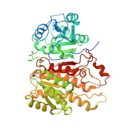
Crystal structure of hexanoyl-CoA bound to beta-ketoacyl reductase FabG4 of Mycobacterium tuberculosis

Dutta, D.Bhattacharyya, S.Roychowdhury, A.Biswas, R.Das, A.K.

(2013) Biochem J 450: 127-139

  • DOI: https://doi.org/10.1042/BJ20121107
  • Primary Citation of Related Structures:  
    3V1U, 4FW8

  • PubMed Abstract: 

    FabGs, or β-oxoacyl reductases, are involved in fatty acid synthesis. The reaction entails NADPH/NADH-mediated conversion of β-oxoacyl-ACP (acyl-carrier protein) into β-hydroxyacyl-ACP. HMwFabGs (high-molecular-weight FabG) form a phylogenetically separate group of FabG enzymes. FabG4, an HMwFabG from Mycobacterium tuberculosis, contains two distinct domains, an N-terminal 'flavodoxintype' domain and a C-terminal oxoreductase domain. The catalytically active C-terminal domain utilizes NADH to reduce β-oxoacyl-CoA to β-hydroxyacyl-CoA. In the present study the crystal structures of the FabG4-NADH binary complex and the FabG4-NAD+-hexanoyl-CoA ternary complex have been determined to understand the substrate specificity and catalytic mechanism of FabG4. This is the first report to demonstrate how FabG4 interacts with its coenzyme NADH and hexanoyl-CoA that mimics an elongating fattyacyl chain covalently linked with CoA. Structural analysis shows that the binding of hexanoyl-CoA within the active site cavity of FabG significantly differs from that of the C16 fattyacyl substrate bound to mycobacterial FabI [InhA (enoyl-ACP reductase)]. The ternary complex reveals that both loop I and loop II interact with the phosphopantetheine moiety of CoA or ACP to align the covalently linked fattyacyl substrate near the active site. Structural data ACP inhibition studies indicate that FabG4 can accept both CoA- and ACP-based fattyacyl substrates. We have also shown that in the FabG4 dimer Arg146 and Arg445 of one monomer interact with the C-terminus of the second monomer to play pivotal role in substrate association and catalysis.


  • Organizational Affiliation

    Department of Biotechnology, Indian Institute of Technology Kharagpur, Kharagpur 721302, India.


Macromolecules
Find similar proteins by:  (by identity cutoff)  |  3D Structure
Entity ID: 1
MoleculeChains Sequence LengthOrganismDetailsImage
3-oxoacyl-(Acyl-carrier-protein) reductase462Mycobacterium tuberculosis H37RvMutation(s): 0 
Gene Names: fabG-1fabG4MT0256Rv0242c
EC: 1.1.1.100
UniProt
Find proteins for O53665 (Mycobacterium tuberculosis (strain CDC 1551 / Oshkosh))
Explore O53665 
Go to UniProtKB:  O53665
Entity Groups  
Sequence Clusters30% Identity50% Identity70% Identity90% Identity95% Identity100% Identity
UniProt GroupO53665
Sequence Annotations
Expand
  • Reference Sequence
Small Molecules
Ligands 3 Unique
IDChains Name / Formula / InChI Key2D Diagram3D Interactions
HXC
Query on HXC

Download Ideal Coordinates CCD File 
C [auth A]HEXANOYL-COENZYME A
C27 H46 N7 O17 P3 S
OEXFMSFODMQEPE-HDRQGHTBSA-N
NAD
Query on NAD

Download Ideal Coordinates CCD File 
B [auth A]NICOTINAMIDE-ADENINE-DINUCLEOTIDE
C21 H27 N7 O14 P2
BAWFJGJZGIEFAR-NNYOXOHSSA-N
ZPG
Query on ZPG

Download Ideal Coordinates CCD File 
D [auth A](2S,5R,8R,11S,14S,17S,21R)-5,8,11,14,17-PENTAMETHYL-4,7,10,13,16,19-HEXAOXADOCOSANE-2,21-DIOL
C21 H44 O8
XGDXHSHOQKNIEW-YSPFXGIPSA-N
Experimental Data & Validation

Experimental Data

  • Method: X-RAY DIFFRACTION
  • Resolution: 2.50 Å
  • R-Value Free: 0.240 
  • R-Value Work: 0.190 
  • R-Value Observed: 0.192 
  • Space Group: P 32 2 1
Unit Cell:
Length ( Å )Angle ( ˚ )
a = 103.059α = 90
b = 103.059β = 90
c = 76.047γ = 120
Software Package:
Software NamePurpose
SCALAdata scaling
PHASERphasing
REFMACrefinement
PDB_EXTRACTdata extraction
XSCALEdata scaling

Structure Validation

View Full Validation Report


Ligand Structure Quality Assessment 


Entry History 

Deposition Data

Revision History  (Full details and data files)

  • Version 1.0: 2012-11-28
    Type: Initial release
  • Version 1.1: 2012-12-05
    Changes: Database references
  • Version 1.2: 2014-03-12
    Changes: Database references
  • Version 1.3: 2023-11-08
    Changes: Data collection, Database references, Derived calculations, Refinement description