3UOT

Crystal Structure of MDC1 FHA Domain in Complex with a Phosphorylated Peptide from the MDC1 N-terminus


Experimental Data Snapshot

  • Method: X-RAY DIFFRACTION
  • Resolution: 1.80 Å
  • R-Value Free: 0.243 
  • R-Value Work: 0.205 
  • R-Value Observed: 0.207 

wwPDB Validation   3D Report Full Report


This is version 1.1 of the entry. See complete history


Literature

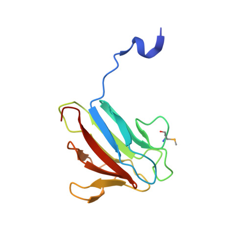
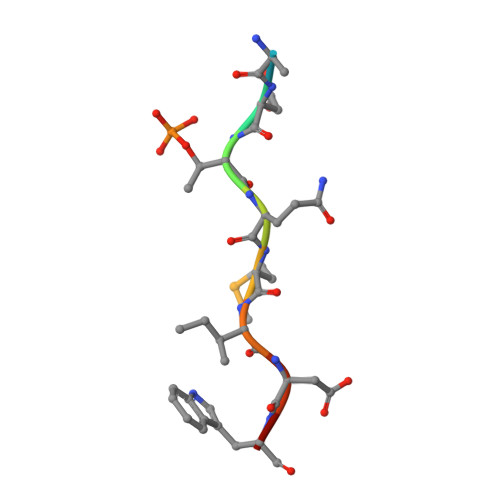
The molecular basis of ATM-dependent dimerization of the Mdc1 DNA damage checkpoint mediator.

Jungmichel, S.Clapperton, J.A.Lloyd, J.Hari, F.J.Spycher, C.Pavic, L.Li, J.Haire, L.F.Bonalli, M.Larsen, D.H.Lukas, C.Lukas, J.MacMillan, D.Nielsen, M.L.Stucki, M.Smerdon, S.J.

(2012) Nucleic Acids Res 40: 3913-3928

  • DOI: https://doi.org/10.1093/nar/gkr1300
  • Primary Citation of Related Structures:  
    3UN0, 3UOT

  • PubMed Abstract: 

    Mdc1 is a large modular phosphoprotein scaffold that maintains signaling and repair complexes at double-stranded DNA break sites. Mdc1 is anchored to damaged chromatin through interaction of its C-terminal BRCT-repeat domain with the tail of γH2AX following DNA damage, but the role of the N-terminal forkhead-associated (FHA) domain remains unclear. We show that a major binding target of the Mdc1 FHA domain is a previously unidentified DNA damage and ATM-dependent phosphorylation site near the N-terminus of Mdc1 itself. Binding to this motif stabilizes a weak self-association of the FHA domain to form a tight dimer. X-ray structures of free and complexed Mdc1 FHA domain reveal a 'head-to-tail' dimerization mechanism that is closely related to that seen in pre-activated forms of the Chk2 DNA damage kinase, and which both positively and negatively influences Mdc1 FHA domain-mediated interactions in human cells prior to and following DNA damage.


  • Organizational Affiliation

    Institute of Veterinary Biochemistry and Molecular Biology, University of Zürich - Irchel, Winterthurerstrasse 190, CH-8057 Zürich, Switzerland.


Macromolecules
Find similar proteins by:  (by identity cutoff)  |  3D Structure
Entity ID: 1
MoleculeChains Sequence LengthOrganismDetailsImage
Mediator of DNA damage checkpoint protein 1
A, B
122Homo sapiensMutation(s): 0 
Gene Names: MDC1KIAA0170NFBD1
UniProt & NIH Common Fund Data Resources
Find proteins for Q14676 (Homo sapiens)
Explore Q14676 
Go to UniProtKB:  Q14676
PHAROS:  Q14676
GTEx:  ENSG00000137337 
Entity Groups  
Sequence Clusters30% Identity50% Identity70% Identity90% Identity95% Identity100% Identity
UniProt GroupQ14676
Sequence Annotations
Expand
  • Reference Sequence

Find similar proteins by:  Sequence   |   3D Structure  

Entity ID: 2
MoleculeChains Sequence LengthOrganismDetailsImage
Mediator of DNA damage checkpoint protein 1C [auth D],
D [auth E]
10Homo sapiensMutation(s): 1 
UniProt & NIH Common Fund Data Resources
Find proteins for Q14676 (Homo sapiens)
Explore Q14676 
Go to UniProtKB:  Q14676
PHAROS:  Q14676
GTEx:  ENSG00000137337 
Entity Groups  
Sequence Clusters30% Identity50% Identity70% Identity90% Identity95% Identity100% Identity
UniProt GroupQ14676
Sequence Annotations
Expand
  • Reference Sequence
Small Molecules
Modified Residues  2 Unique
IDChains TypeFormula2D DiagramParent
MSE
Query on MSE
A, B
L-PEPTIDE LINKINGC5 H11 N O2 SeMET
TPO
Query on TPO
C [auth D],
D [auth E]
L-PEPTIDE LINKINGC4 H10 N O6 PTHR
Experimental Data & Validation

Experimental Data

  • Method: X-RAY DIFFRACTION
  • Resolution: 1.80 Å
  • R-Value Free: 0.243 
  • R-Value Work: 0.205 
  • R-Value Observed: 0.207 
  • Space Group: P 21 21 21
Unit Cell:
Length ( Å )Angle ( ˚ )
a = 58.485α = 90
b = 59.88β = 90
c = 72.081γ = 90
Software Package:
Software NamePurpose
HKL-2000data collection
SOLVEphasing
REFMACrefinement
HKL-2000data reduction
SCALEPACKdata scaling

Structure Validation

View Full Validation Report


Entry History 

Deposition Data

Revision History  (Full details and data files)

  • Version 1.0: 2011-12-28
    Type: Initial release
  • Version 1.1: 2012-07-18
    Changes: Database references