3U2B

Structure of the Sox4 HMG domain bound to DNA


Experimental Data Snapshot

  • Method: X-RAY DIFFRACTION
  • Resolution: 2.40 Å
  • R-Value Free: 0.280 
  • R-Value Work: 0.235 
  • R-Value Observed: 0.238 

wwPDB Validation   3D Report Full Report


This is version 1.2 of the entry. See complete history


Literature

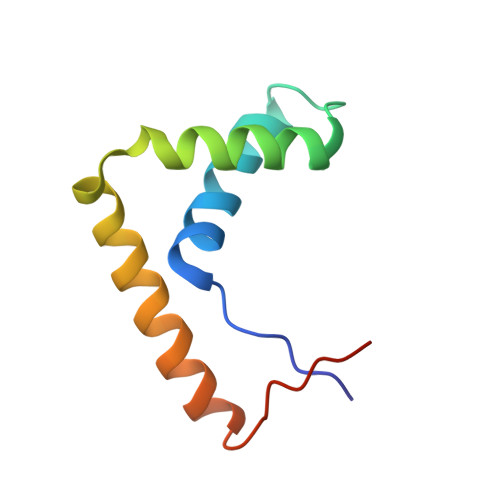
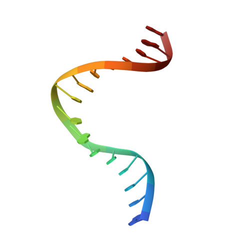
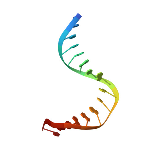
The crystal structure of the Sox4 HMG domain-DNA complex suggests a mechanism for positional interdependence in DNA recognition

Jauch, R.Ng, C.K.L.Narasimhan, K.Kolatkar, P.R.

(2012) Biochem J 443: 39-47

  • DOI: https://doi.org/10.1042/BJ20111768
  • Primary Citation of Related Structures:  
    3U2B

  • PubMed Abstract: 

    It has recently been proposed that the sequence preferences of DNA-binding TFs (transcription factors) can be well described by models that include the positional interdependence of the nucleotides of the target sites. Such binding models allow for multiple motifs to be invoked, such as principal and secondary motifs differing at two or more nucleotide positions. However, the structural mechanisms underlying the accommodation of such variant motifs by TFs remain elusive. In the present study we examine the crystal structure of the HMG (high-mobility group) domain of Sox4 [Sry (sex-determining region on the Y chromosome)-related HMG box 4] bound to DNA. By comparing this structure with previously solved structures of Sox17 and Sox2, we observed subtle conformational differences at the DNA-binding interface. Furthermore, using quantitative electrophoretic mobility-shift assays we validated the positional interdependence of two nucleotides and the presence of a secondary Sox motif in the affinity landscape of Sox4. These results suggest that a concerted rearrangement of two interface amino acids enables Sox4 to accommodate primary and secondary motifs. The structural adaptations lead to altered dinucleotide preferences that mutually reinforce each other. These analyses underline the complexity of the DNA recognition by TFs and provide an experimental validation for the conceptual framework of positional interdependence and secondary binding motifs.


  • Organizational Affiliation

    Laboratory for Structural Biochemistry, Genome Institute of Singapore, 60 Biopolis St, Singapore 138672. jauchr@gis.a-star.edu.sg


Macromolecules

Find similar proteins by:  (by identity cutoff)  |  3D Structure
Entity ID: 3
MoleculeChains Sequence LengthOrganismDetailsImage
Transcription factor SOX-479Mus musculusMutation(s): 0 
Gene Names: Sox4Sox-4
UniProt & NIH Common Fund Data Resources
Find proteins for Q06831 (Mus musculus)
Explore Q06831 
Go to UniProtKB:  Q06831
IMPC:  MGI:98366
Entity Groups  
Sequence Clusters30% Identity50% Identity70% Identity90% Identity95% Identity100% Identity
UniProt GroupQ06831
Sequence Annotations
Expand
  • Reference Sequence

Find similar nucleic acids by:  Sequence   |   3D Structure  

Entity ID: 1
MoleculeChains LengthOrganismImage
DNA (5'-D(*GP*TP*CP*TP*CP*TP*AP*TP*TP*GP*TP*CP*CP*TP*GP*G)-3')16N/A
Sequence Annotations
Expand
  • Reference Sequence

Find similar nucleic acids by:  Sequence   |   3D Structure  

Entity ID: 2
MoleculeChains LengthOrganismImage
DNA (5'-D(*CP*CP*AP*GP*GP*AP*CP*AP*AP*TP*AP*GP*AP*GP*AP*C)-3')16N/A
Sequence Annotations
Expand
  • Reference Sequence
Experimental Data & Validation

Experimental Data

  • Method: X-RAY DIFFRACTION
  • Resolution: 2.40 Å
  • R-Value Free: 0.280 
  • R-Value Work: 0.235 
  • R-Value Observed: 0.238 
  • Space Group: P 32 2 1
Unit Cell:
Length ( Å )Angle ( ˚ )
a = 69.941α = 90
b = 69.941β = 90
c = 63.205γ = 120
Software Package:
Software NamePurpose
HKL-2000data collection
PHASESphasing
PHENIXrefinement
HKL-2000data reduction
HKL-2000data scaling

Structure Validation

View Full Validation Report


Entry History 

Deposition Data

Revision History  (Full details and data files)

  • Version 1.0: 2011-12-28
    Type: Initial release
  • Version 1.1: 2014-03-12
    Changes: Database references
  • Version 1.2: 2024-03-20
    Changes: Data collection, Database references