3TOW

Crystal Structure of the MABP Domain of MVB12B of Human ESCRT-I Complex


Experimental Data Snapshot

  • Method: X-RAY DIFFRACTION
  • Resolution: 1.34 Å
  • R-Value Free: 0.193 
  • R-Value Work: 0.165 
  • R-Value Observed: 0.166 

wwPDB Validation   3D Report Full Report


This is version 1.2 of the entry. See complete history


Literature

Structural basis for membrane targeting by the MVB12-associated beta-prism domain of the human ESCRT-I MVB12 subunit.

Boura, E.Hurley, J.H.

(2012) Proc Natl Acad Sci U S A 109: 1901-1906

  • DOI: https://doi.org/10.1073/pnas.1117597109
  • Primary Citation of Related Structures:  
    3TOW

  • PubMed Abstract: 

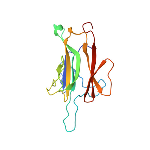
    MVB12-associated β-prism (MABP) domains are predicted to occur in a diverse set of membrane-associated bacterial and eukaryotic proteins, but their existence, structure, and biochemical properties have not been characterized experimentally. Here, we find that the MABP domains of the MVB12A and B subunits of ESCRT-I are functional modules that bind in vitro to liposomes containing acidic lipids depending on negative charge density. The MABP domain is capable of autonomously localizing to subcellular puncta and to the plasma membrane. The 1.3-Å atomic resolution crystal structure of the MVB12B MABP domain reveals a β-prism fold, a hydrophobic membrane-anchoring loop, and an electropositive phosphoinositide-binding patch. The basic patch is open, which explains how it senses negative charge density but lacks stereoselectivity. These observations show how ESCRT-I could act as a coincidence detector for acidic phospholipids and protein ligands, enabling it to function both in protein transport at endosomes and in cytokinesis and viral budding at the plasma membrane.


  • Organizational Affiliation

    Laboratory of Molecular Biology, National Institute of Diabetes and Digestive and Kidney Diseases, National Institutes of Health, Bethesda, MD 20892, USA.


Macromolecules
Find similar proteins by:  (by identity cutoff)  |  3D Structure
Entity ID: 1
MoleculeChains Sequence LengthOrganismDetailsImage
Multivesicular body subunit 12B152Homo sapiensMutation(s): 0 
Gene Names: C9orf28FAM125BMVB12B
Membrane Entity: Yes 
UniProt & NIH Common Fund Data Resources
Find proteins for Q9H7P6 (Homo sapiens)
Explore Q9H7P6 
Go to UniProtKB:  Q9H7P6
PHAROS:  Q9H7P6
GTEx:  ENSG00000196814 
Entity Groups  
Sequence Clusters30% Identity50% Identity70% Identity90% Identity95% Identity100% Identity
UniProt GroupQ9H7P6
Sequence Annotations
Expand
  • Reference Sequence
Experimental Data & Validation

Experimental Data

  • Method: X-RAY DIFFRACTION
  • Resolution: 1.34 Å
  • R-Value Free: 0.193 
  • R-Value Work: 0.165 
  • R-Value Observed: 0.166 
  • Space Group: P 21 21 21
Unit Cell:
Length ( Å )Angle ( ˚ )
a = 32.371α = 90
b = 52.607β = 90
c = 71.388γ = 90
Software Package:
Software NamePurpose
MAR345dtbdata collection
SHELXSphasing
PHENIXrefinement
HKL-2000data reduction
HKL-2000data scaling

Structure Validation

View Full Validation Report


Entry History 

Deposition Data

Revision History  (Full details and data files)

  • Version 1.0: 2012-01-11
    Type: Initial release
  • Version 1.1: 2013-11-27
    Changes: Database references
  • Version 1.2: 2024-02-28
    Changes: Data collection, Database references