3TJQ

N-domain of HtrA1


Experimental Data Snapshot

  • Method: X-RAY DIFFRACTION
  • Resolution: 2.00 Å
  • R-Value Free: 0.270 
  • R-Value Work: 0.217 
  • R-Value Observed: 0.222 

wwPDB Validation   3D Report Full Report


This is version 1.2 of the entry. See complete history


Literature

Structural and Functional Analysis of HtrA1 and Its Subdomains.

Eigenbrot, C.Ultsch, M.Lipari, M.T.Moran, P.Lin, S.J.Ganesan, R.Quan, C.Tom, J.Sandoval, W.van Lookeren Campagne, M.Kirchhofer, D.

(2012) Structure 20: 1040-1050

  • DOI: https://doi.org/10.1016/j.str.2012.03.021
  • Primary Citation of Related Structures:  
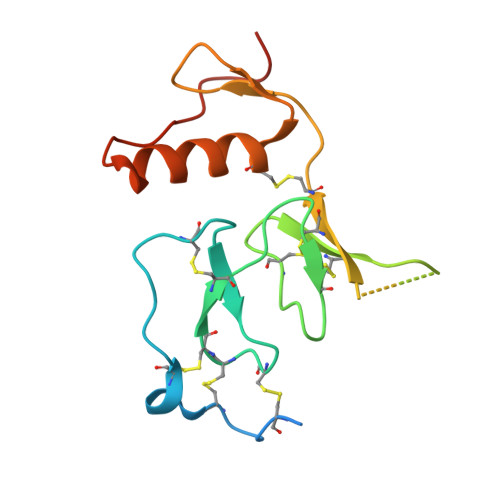
    3TJN, 3TJO, 3TJQ

  • PubMed Abstract: 

    The homotrimeric human serine protease HtrA1 is homologous to bacterial HtrA proteases regarding the trypsin-like catalytic and PDZ domains but differs by the presence of an N-terminal domain with IGFBP and Kazal homology. The crystal structures and SAXS analysis presented herein reveal the rare tandem of IGFBP- and Kazal-like modules, a protease active site that adopts a competent conformation in the absence of substrate or inhibitor and a model for the intact protein in solution. Highly sensitive enzymatic assays and binding studies demonstrate that the N-terminal tandem has no apparent effect on protease activity, and in accordance with the structure-based predictions, neither the IGFBP- nor Kazal-like module retains the function of their prototype proteins. Our structures of the unliganded HtrA1 active site suggest two-state equilibrium and a "conformational selection" model, in which substrate binds to the active conformer.


  • Organizational Affiliation

    Department of Structural Biology, Genentech, Inc., South San Francisco, CA 94080, USA.


Macromolecules
Find similar proteins by:  (by identity cutoff)  |  3D Structure
Entity ID: 1
MoleculeChains Sequence LengthOrganismDetailsImage
Serine protease HTRA1140Homo sapiensMutation(s): 0 
Gene Names: HTRAHTRA1PRSS11
EC: 3.4.21
UniProt & NIH Common Fund Data Resources
Find proteins for Q92743 (Homo sapiens)
Explore Q92743 
Go to UniProtKB:  Q92743
PHAROS:  Q92743
GTEx:  ENSG00000166033 
Entity Groups  
Sequence Clusters30% Identity50% Identity70% Identity90% Identity95% Identity100% Identity
UniProt GroupQ92743
Sequence Annotations
Expand
  • Reference Sequence
Small Molecules
Ligands 4 Unique
IDChains Name / Formula / InChI Key2D Diagram3D Interactions
PT
Query on PT

Download Ideal Coordinates CCD File 
B [auth A],
F [auth A]
PLATINUM (II) ION
Pt
HRGDZIGMBDGFTC-UHFFFAOYSA-N
GOL
Query on GOL

Download Ideal Coordinates CCD File 
J [auth A]GLYCEROL
C3 H8 O3
PEDCQBHIVMGVHV-UHFFFAOYSA-N
SCN
Query on SCN

Download Ideal Coordinates CCD File 
I [auth A],
L [auth A],
M [auth A],
N [auth A]
THIOCYANATE ION
C N S
ZMZDMBWJUHKJPS-UHFFFAOYSA-M
CL
Query on CL

Download Ideal Coordinates CCD File 
C [auth A]
D [auth A]
E [auth A]
G [auth A]
H [auth A]
C [auth A],
D [auth A],
E [auth A],
G [auth A],
H [auth A],
K [auth A]
CHLORIDE ION
Cl
VEXZGXHMUGYJMC-UHFFFAOYSA-M
Experimental Data & Validation

Experimental Data

  • Method: X-RAY DIFFRACTION
  • Resolution: 2.00 Å
  • R-Value Free: 0.270 
  • R-Value Work: 0.217 
  • R-Value Observed: 0.222 
  • Space Group: P 31 2 1
Unit Cell:
Length ( Å )Angle ( ˚ )
a = 72.085α = 90
b = 72.085β = 90
c = 41.289γ = 120
Software Package:
Software NamePurpose
BOSdata collection
PHENIXmodel building
REFMACrefinement
HKL-2000data reduction
HKL-2000data scaling
PHENIXphasing

Structure Validation

View Full Validation Report


Entry History 

Deposition Data

Revision History  (Full details and data files)

  • Version 1.0: 2012-05-16
    Type: Initial release
  • Version 1.1: 2012-06-13
    Changes: Database references
  • Version 1.2: 2012-06-27
    Changes: Database references