3SME

Structure of PTP1B inactivated by H2O2/bicarbonate


Experimental Data Snapshot

  • Method: X-RAY DIFFRACTION
  • Resolution: 1.70 Å
  • R-Value Free: 0.210 
  • R-Value Work: 0.197 
  • R-Value Observed: 0.198 

wwPDB Validation   3D Report Full Report


This is version 1.1 of the entry. See complete history


Literature

The Biological Buffer Bicarbonate/CO(2) Potentiates H(2)O(2)-Mediated Inactivation of Protein Tyrosine Phosphatases.

Zhou, H.Singh, H.Parsons, Z.D.Lewis, S.M.Bhattacharya, S.Seiner, D.R.Labutti, J.N.Reilly, T.J.Tanner, J.J.Gates, K.S.

(2011) J Am Chem Soc 133: 15803-15805

  • DOI: https://doi.org/10.1021/ja2077137
  • Primary Citation of Related Structures:  
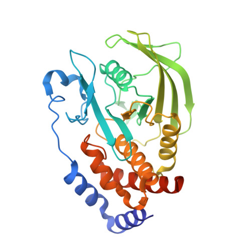
    3SME

  • PubMed Abstract: 

    Hydrogen peroxide is a cell signaling agent that inactivates protein tyrosine phosphatases (PTPs) via oxidation of their catalytic cysteine residue. PTPs are inactivated rapidly during H(2)O(2)-mediated cellular signal transduction processes, but, paradoxically, hydrogen peroxide is a rather sluggish PTP inactivator in vitro. Here we present evidence that the biological buffer bicarbonate/CO(2) potentiates the ability of H(2)O(2) to inactivate PTPs. The results of biochemical experiments and high-resolution crystallographic analysis are consistent with a mechanism involving oxidation of the catalytic cysteine residue by peroxymonocarbonate generated via the reaction of H(2)O(2) with HCO(3)(-)/CO(2).


  • Organizational Affiliation

    Department of Chemistry, University of Missouri, Columbia, Missouri 65211, United States.


Macromolecules
Find similar proteins by:  (by identity cutoff)  |  3D Structure
Entity ID: 1
MoleculeChains Sequence LengthOrganismDetailsImage
Tyrosine-protein phosphatase non-receptor type 1300Homo sapiensMutation(s): 0 
Gene Names: PTP1BPTPN1
EC: 3.1.3.48
UniProt & NIH Common Fund Data Resources
Find proteins for P18031 (Homo sapiens)
Explore P18031 
Go to UniProtKB:  P18031
PHAROS:  P18031
GTEx:  ENSG00000196396 
Entity Groups  
Sequence Clusters30% Identity50% Identity70% Identity90% Identity95% Identity100% Identity
UniProt GroupP18031
Sequence Annotations
Expand
  • Reference Sequence
Small Molecules
Ligands 1 Unique
IDChains Name / Formula / InChI Key2D Diagram3D Interactions
MG
Query on MG

Download Ideal Coordinates CCD File 
B [auth A]MAGNESIUM ION
Mg
JLVVSXFLKOJNIY-UHFFFAOYSA-N
Experimental Data & Validation

Experimental Data

  • Method: X-RAY DIFFRACTION
  • Resolution: 1.70 Å
  • R-Value Free: 0.210 
  • R-Value Work: 0.197 
  • R-Value Observed: 0.198 
  • Space Group: P 31 2 1
Unit Cell:
Length ( Å )Angle ( ˚ )
a = 88α = 90
b = 88β = 90
c = 104γ = 120
Software Package:
Software NamePurpose
d*TREKdata reduction
PHENIXrefinement
PDB_EXTRACTdata extraction
d*TREKdata scaling
PHENIXphasing

Structure Validation

View Full Validation Report


Entry History 

Deposition Data

Revision History  (Full details and data files)

  • Version 1.0: 2011-10-19
    Type: Initial release
  • Version 1.1: 2023-09-13
    Changes: Data collection, Database references, Derived calculations, Refinement description