Structure and Dynamics of a Stabilized Coiled-Coil Domain in the P-TEFb Regulator Hexim1.
Bigalke, J.M., Dames, S.A., Blankenfeldt, W., Grzesiek, S., Geyer, M.(2011) J Mol Biol 414: 639-653
- PubMed: 22033481
- DOI: https://doi.org/10.1016/j.jmb.2011.10.022
- Primary Citation of Related Structures:
3S9G - PubMed Abstract:
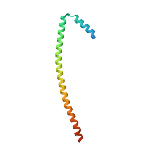
The positive transcription elongation factor P-TEFb mediates the transition from transcription initiation to productive elongation by phosphorylation of the C-terminal domain of RNA polymerase II. P-TEFb is negatively regulated by the cellular protein Hexim1 (hexamethylene bisacetamide-inducible protein 1), which is highly conserved in higher eukaryotes. The C-terminal coiled-coil domain of Hexim1 recognizes the Cyclin T subunit of P-TEFb, whereas a central PYNT motif is required to inhibit the cyclin-dependent kinase Cdk9 by a yet unknown mechanism. Here, the crystal structure of the Cyclin T-binding domain (TBD) of human Hexim1 was determined at 2.1 Å resolution using a deletion mutant of three residues in its central stammer motif. The structure showed a continuous parallel coiled-coil domain of nine hepta-repeats with a preceding helix encompassing up to 15 residues. Two uncommon residues at heptad a positions in the N-terminal part of the coiled-coil structure, Lys284 and Tyr291, stabilize the preceding helix by a tight intermolecular hydrogen bond network with residues of the opposing chain. These interactions delineate a characteristic turn between both helices that is supposed to mediate binding to Cyclin T1. Stabilization of the coiled-coil domain by deletion of the stammer region was confirmed by NMR spectroscopic and backbone dynamic analyses analyzing wild-type TBD and three mutant variants. This study thus provides structural insights into the recognition of the regulator protein Hexim1 by P-TEFb and the modulation of coiled-coil dynamics by specific discontinuities.
Organizational Affiliation:
Max-Planck-Institut für molekulare Physiologie, Abteilung Physikalische Biochemie, Otto-Hahn-Strasse 11, 44227 Dortmund, Germany.