3S6W

Crystal structure of Tudor domain of human TDRD3


Experimental Data Snapshot

  • Method: X-RAY DIFFRACTION
  • Resolution: 1.78 Å
  • R-Value Free: 0.206 
  • R-Value Work: 0.183 
  • R-Value Observed: 0.184 

wwPDB Validation   3D Report Full Report


This is version 1.3 of the entry. See complete history


Literature

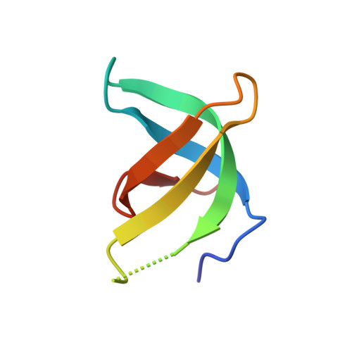
Crystal structure of TDRD3 and methyl-arginine binding characterization of TDRD3, SMN and SPF30

Liu, K.Guo, Y.H.Liu, H.P.Bian, C.B.Lam, R.Liu, Y.Mackenzie, F.Rojas, L.A.Reinberg, D.Bedford, M.T.Xu, R.M.Min, J.R.

(2012) PLoS One 7: e30375-e30375

  • DOI: https://doi.org/10.1371/journal.pone.0030375
  • Primary Citation of Related Structures:  
    3PMT, 3S6W

  • PubMed Abstract: 

    SMN (Survival motor neuron protein) was characterized as a dimethyl-arginine binding protein over ten years ago. TDRD3 (Tudor domain-containing protein 3) and SPF30 (Splicing factor 30 kDa) were found to bind to various methyl-arginine proteins including Sm proteins as well later on. Recently, TDRD3 was shown to be a transcriptional coactivator, and its transcriptional activity is dependent on its ability to bind arginine-methylated histone marks. In this study, we systematically characterized the binding specificity and affinity of the Tudor domains of these three proteins quantitatively. Our results show that TDRD3 preferentially recognizes asymmetrical dimethylated arginine mark, and SMN is a very promiscuous effector molecule, which recognizes different arginine containing sequence motifs and preferentially binds symmetrical dimethylated arginine. SPF30 is the weakest methyl-arginine binder, which only binds the GAR motif sequences in our library. In addition, we also reported high-resolution crystal structures of the Tudor domain of TDRD3 in complex with two small molecules, which occupy the aromatic cage of TDRD3.


  • Organizational Affiliation

    Hubei Key Laboratory of Genetic Regulation and Integrative Biology, College of Life Science, Huazhong Normal University, Wuhan, People's Republic of China.


Macromolecules
Find similar proteins by:  (by identity cutoff)  |  3D Structure
Entity ID: 1
MoleculeChains Sequence LengthOrganismDetailsImage
Tudor domain-containing protein 354Homo sapiensMutation(s): 0 
Gene Names: TDRD3
UniProt & NIH Common Fund Data Resources
Find proteins for Q9H7E2 (Homo sapiens)
Explore Q9H7E2 
Go to UniProtKB:  Q9H7E2
PHAROS:  Q9H7E2
GTEx:  ENSG00000083544 
Entity Groups  
Sequence Clusters30% Identity50% Identity70% Identity90% Identity95% Identity100% Identity
UniProt GroupQ9H7E2
Sequence Annotations
Expand
  • Reference Sequence
Experimental Data & Validation

Experimental Data

  • Method: X-RAY DIFFRACTION
  • Resolution: 1.78 Å
  • R-Value Free: 0.206 
  • R-Value Work: 0.183 
  • R-Value Observed: 0.184 
  • Space Group: P 65
Unit Cell:
Length ( Å )Angle ( ˚ )
a = 41.93α = 90
b = 41.93β = 90
c = 61.478γ = 120
Software Package:
Software NamePurpose
MOLREPphasing
REFMACrefinement
PDB_EXTRACTdata extraction
d*TREKdata scaling
DENZOdata reduction
SCALEPACKdata scaling
HKL-2000data scaling

Structure Validation

View Full Validation Report


Entry History 

Deposition Data

Revision History  (Full details and data files)

  • Version 1.0: 2012-03-07
    Type: Initial release
  • Version 1.1: 2012-05-02
    Changes: Database references
  • Version 1.2: 2017-11-08
    Changes: Refinement description
  • Version 1.3: 2023-11-01
    Changes: Data collection, Database references, Derived calculations, Refinement description