3RW9

Crystal Structure of human Spermidine Synthase in Complex with decarboxylated S-adenosylhomocysteine


Experimental Data Snapshot

  • Method: X-RAY DIFFRACTION
  • Resolution: 2.00 Å
  • R-Value Free: 0.259 
  • R-Value Work: 0.211 
  • R-Value Observed: 0.213 

wwPDB Validation   3D Report Full Report


Ligand Structure Quality Assessment 


This is version 1.2 of the entry. See complete history


Literature

Binding and inhibition of human spermidine synthase by decarboxylated S-adenosylhomocysteine.

Seckute, J.McCloskey, D.E.Thomas, H.J.Secrist, J.A.Pegg, A.E.Ealick, S.E.

(2011) Protein Sci 20: 1836-1844

  • DOI: https://doi.org/10.1002/pro.717
  • Primary Citation of Related Structures:  
    3RW9

  • PubMed Abstract: 

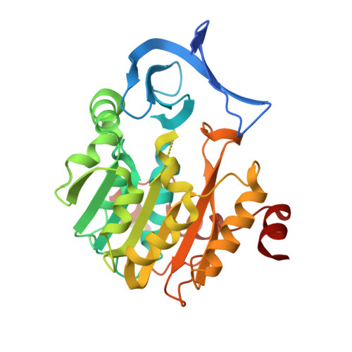
    Aminopropyltransferases are essential enzymes that form polyamines in eukaryotic and most prokaryotic cells. Spermidine synthase (SpdS) is one of the most well-studied enzymes in this biosynthetic pathway. The enzyme uses decarboxylated S-adenosylmethionine and a short-chain polyamine (putrescine) to make a medium-chain polyamine (spermidine) and 5'-deoxy-5'-methylthioadenosine as a byproduct. Here, we report a new spermidine synthase inhibitor, decarboxylated S-adenosylhomocysteine (dcSAH). The inhibitor was synthesized, and dose-dependent inhibition of human, Thermatoga maritima, and Plasmodium falciparum spermidine synthases, as well as functionally homologous human spermine synthase, was determined. The human SpdS/dcSAH complex structure was determined by X-ray crystallography at 2.0 Å resolution and showed consistent active site positioning and coordination with previously known structures. Isothermal calorimetry binding assays confirmed inhibitor binding to human SpdS with K(d) of 1.1 ± 0.3 μM in the absence of putrescine and 3.2 ± 0.1 μM in the presence of putrescine. These results indicate a potential for further inhibitor development based on the dcSAH scaffold.


  • Organizational Affiliation

    Department of Chemistry and Chemical Biology, Cornell University, Ithaca, New York 14853, USA.


Macromolecules
Find similar proteins by:  (by identity cutoff)  |  3D Structure
Entity ID: 1
MoleculeChains Sequence LengthOrganismDetailsImage
Spermidine synthase
A, B
304Homo sapiensMutation(s): 0 
Gene Names: SRMSPS1SRML1
EC: 2.5.1.16
UniProt & NIH Common Fund Data Resources
Find proteins for P19623 (Homo sapiens)
Explore P19623 
Go to UniProtKB:  P19623
PHAROS:  P19623
GTEx:  ENSG00000116649 
Entity Groups  
Sequence Clusters30% Identity50% Identity70% Identity90% Identity95% Identity100% Identity
UniProt GroupP19623
Sequence Annotations
Expand
  • Reference Sequence
Small Molecules
Ligands 1 Unique
IDChains Name / Formula / InChI Key2D Diagram3D Interactions
DSH
Query on DSH

Download Ideal Coordinates CCD File 
C [auth A],
D [auth B]
5'-S-(3-aminopropyl)-5'-thioadenosine
C13 H20 N6 O3 S
FUSRAALGPJJIRO-QYVSTXNMSA-N
Binding Affinity Annotations 
IDSourceBinding Affinity
DSH Binding MOAD:  3RW9 Kd: 1100 (nM) from 1 assay(s)
PDBBind:  3RW9 Kd: 1100 (nM) from 1 assay(s)
Experimental Data & Validation

Experimental Data

  • Method: X-RAY DIFFRACTION
  • Resolution: 2.00 Å
  • R-Value Free: 0.259 
  • R-Value Work: 0.211 
  • R-Value Observed: 0.213 
  • Space Group: P 1 21 1
Unit Cell:
Length ( Å )Angle ( ˚ )
a = 58.177α = 90
b = 60.816β = 108.05
c = 87.021γ = 90
Software Package:
Software NamePurpose
REFMACrefinement
PDB_EXTRACTdata extraction
ADSCdata collection
HKL-2000data reduction
HKL-2000data scaling
CNSphasing

Structure Validation

View Full Validation Report


Ligand Structure Quality Assessment 


Entry History 

Deposition Data

Revision History  (Full details and data files)

  • Version 1.0: 2011-09-21
    Type: Initial release
  • Version 1.1: 2011-11-02
    Changes: Database references
  • Version 1.2: 2024-02-28
    Changes: Data collection, Database references, Derived calculations