3QY9

The Crystal Structure of Dihydrodipicolinate reductase from Staphylococcus aureus


Experimental Data Snapshot

  • Method: X-RAY DIFFRACTION
  • Resolution: 1.80 Å
  • R-Value Free: 0.225 
  • R-Value Work: 0.198 
  • R-Value Observed: 0.199 

wwPDB Validation   3D Report Full Report


This is version 1.2 of the entry. See complete history


Literature

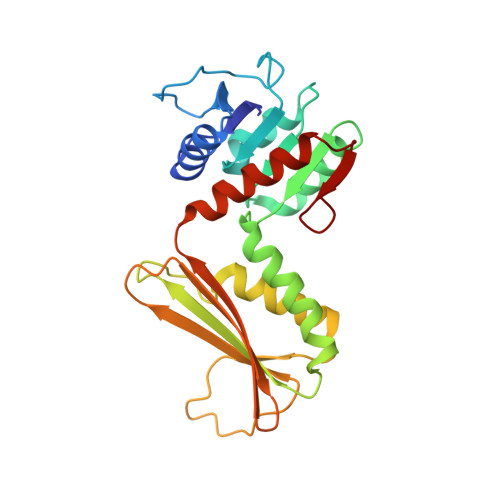
Structure and nucleotide specificity of Staphylococcus aureus dihydrodipicolinate reductase (DapB)

Girish, T.S.Navratna, V.Gopal, B.

(2011) FEBS Lett 585: 2561-2567

  • DOI: https://doi.org/10.1016/j.febslet.2011.07.021
  • Primary Citation of Related Structures:  
    3QY9

  • PubMed Abstract: 

    Lysine biosynthesis proceeds by the nucleotide-dependent reduction of dihydrodipicolinate (DHDP) to tetrahydrodipicolinate (THDP) by dihydrodipicolinate reductase (DHDPR). The S. aureus DHDPR structure reveals different conformational states of this enzyme even in the absence of a substrate or nucleotide-cofactor. Despite lacking a conserved basic residue essential for NADPH interaction, S. aureus DHDPR differs from other homologues as NADPH is a more preferred co-factor than NADH. The structure provides a rationale-Lys35 compensates for the co-factor site mutation. These observations are significant for bi-ligand inhibitor design that relies on ligand-induced conformational changes as well as co-factor specificity for this important drug target.


  • Organizational Affiliation

    Molecular Biophysics Unit, Indian Institute of Science, Bangalore, India.


Macromolecules
Find similar proteins by:  (by identity cutoff)  |  3D Structure
Entity ID: 1
MoleculeChains Sequence LengthOrganismDetailsImage
Dihydrodipicolinate reductase
A, B, C, D
243Staphylococcus aureus subsp. aureus COLMutation(s): 0 
Gene Names: dapB
EC: 1.3.1.26
UniProt
Find proteins for Q5HG24 (Staphylococcus aureus (strain COL))
Explore Q5HG24 
Go to UniProtKB:  Q5HG24
Entity Groups  
Sequence Clusters30% Identity50% Identity70% Identity90% Identity95% Identity100% Identity
UniProt GroupQ5HG24
Sequence Annotations
Expand
  • Reference Sequence
Small Molecules
Ligands 3 Unique
IDChains Name / Formula / InChI Key2D Diagram3D Interactions
SO4
Query on SO4

Download Ideal Coordinates CCD File 
AA [auth D]
BA [auth D]
CA [auth D]
DA [auth D]
E [auth A]
AA [auth D],
BA [auth D],
CA [auth D],
DA [auth D],
E [auth A],
F [auth A],
G [auth A],
H [auth A],
K [auth B],
L [auth B],
M [auth B],
N [auth B],
O [auth B],
P [auth B],
R [auth C],
S [auth C],
T [auth C],
U [auth C],
V [auth C],
W [auth C],
Y [auth D],
Z [auth D]
SULFATE ION
O4 S
QAOWNCQODCNURD-UHFFFAOYSA-L
GOL
Query on GOL

Download Ideal Coordinates CCD File 
I [auth A],
Q [auth B]
GLYCEROL
C3 H8 O3
PEDCQBHIVMGVHV-UHFFFAOYSA-N
ACT
Query on ACT

Download Ideal Coordinates CCD File 
J [auth B],
X [auth D]
ACETATE ION
C2 H3 O2
QTBSBXVTEAMEQO-UHFFFAOYSA-M
Experimental Data & Validation

Experimental Data

  • Method: X-RAY DIFFRACTION
  • Resolution: 1.80 Å
  • R-Value Free: 0.225 
  • R-Value Work: 0.198 
  • R-Value Observed: 0.199 
  • Space Group: P 21 21 21
Unit Cell:
Length ( Å )Angle ( ˚ )
a = 85.9α = 90
b = 112.44β = 90
c = 131.61γ = 90
Software Package:
Software NamePurpose
HKL-2000data collection
PHASERphasing
REFMACrefinement
MOSFLMdata reduction
SCALAdata scaling

Structure Validation

View Full Validation Report


Entry History 

Deposition Data

Revision History  (Full details and data files)

  • Version 1.0: 2011-08-03
    Type: Initial release
  • Version 1.1: 2011-09-28
    Changes: Database references
  • Version 1.2: 2023-11-01
    Changes: Data collection, Database references, Derived calculations, Refinement description