3PH9

Crystal structure of the human anterior gradient protein 3


Experimental Data Snapshot

  • Method: X-RAY DIFFRACTION
  • Resolution: 1.83 Å
  • R-Value Free: 0.233 
  • R-Value Work: 0.177 
  • R-Value Observed: 0.180 

wwPDB Validation   3D Report Full Report


This is version 1.2 of the entry. See complete history


Literature

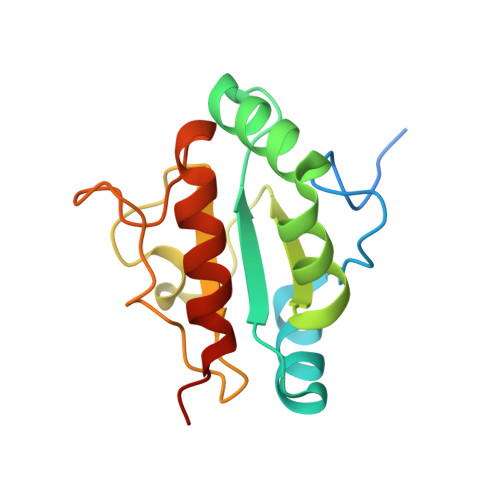
Crystal structure of human anterior gradient protein 3.

Nguyen, V.D.Biterova, E.Salin, M.Wierenga, R.K.Ruddock, L.W.

(2018) Acta Crystallogr F Struct Biol Commun 74: 425-430

  • DOI: https://doi.org/10.1107/S2053230X18009093
  • Primary Citation of Related Structures:  
    3PH9

  • PubMed Abstract: 

    Oxidative protein folding in the endoplasmic reticulum is catalyzed by the protein disulfide isomerase family of proteins. Of the 20 recognized human family members, the structures of eight have been deposited in the PDB along with domains from six more. Three members of this family, ERp18, anterior gradient protein 2 (AGR2) and anterior gradient protein 3 (AGR3), are single-domain proteins which share sequence similarity. While ERp18 has a canonical active-site motif and is involved in native disulfide-bond formation, AGR2 and AGR3 lack elements of the active-site motif found in other family members and may both interact with mucins. In order to better define its function, the structure of AGR3 is required. Here, the recombinant expression, purification, crystallization and crystal structure of human AGR3 are described.


  • Organizational Affiliation

    Faculty of Biochemistry and Molecular Medicine, University of Oulu, Aapistie 7, 90220 Oulu, Finland.


Macromolecules
Find similar proteins by:  (by identity cutoff)  |  3D Structure
Entity ID: 1
MoleculeChains Sequence LengthOrganismDetailsImage
Anterior gradient protein 3 homolog
A, B
151Homo sapiensMutation(s): 0 
Gene Names: AGR3BCMP11UNQ642/PRO1272
EC: 5.3.4.1
UniProt & NIH Common Fund Data Resources
Find proteins for Q8TD06 (Homo sapiens)
Explore Q8TD06 
Go to UniProtKB:  Q8TD06
PHAROS:  Q8TD06
GTEx:  ENSG00000173467 
Entity Groups  
Sequence Clusters30% Identity50% Identity70% Identity90% Identity95% Identity100% Identity
UniProt GroupQ8TD06
Sequence Annotations
Expand
  • Reference Sequence
Experimental Data & Validation

Experimental Data

  • Method: X-RAY DIFFRACTION
  • Resolution: 1.83 Å
  • R-Value Free: 0.233 
  • R-Value Work: 0.177 
  • R-Value Observed: 0.180 
  • Space Group: P 1 21 1
Unit Cell:
Length ( Å )Angle ( ˚ )
a = 33.29α = 90
b = 71.45β = 97.72
c = 59.81γ = 90
Software Package:
Software NamePurpose
ADSCdata collection
MOLREPphasing
REFMACrefinement
XDSdata reduction
XSCALEdata scaling

Structure Validation

View Full Validation Report


Entry History 

Deposition Data

Revision History  (Full details and data files)

  • Version 1.0: 2011-10-19
    Type: Initial release
  • Version 1.1: 2023-07-26
    Changes: Database references
  • Version 1.2: 2023-09-06
    Changes: Data collection, Refinement description