3P8X

Synthesis, Structure, and Biological Activity of des-Side Chain Analogues of 1alpha,25-Dihydroxyvitamin D3 with Substituents at C-18


Experimental Data Snapshot

  • Method: X-RAY DIFFRACTION
  • Resolution: 1.70 Å
  • R-Value Free: 0.209 
  • R-Value Work: 0.189 
  • R-Value Observed: 0.189 

wwPDB Validation   3D Report Full Report


Ligand Structure Quality Assessment 


This is version 1.2 of the entry. See complete history


Literature

Synthesis, Structure, and Biological Activity of des-Side Chain Analogues of 1 ,25-Dihydroxyvitamin D3 with Substituents at C-18

Verlinden, L.Verstuyf, A.Eelen, G.Bouillon, R.Ordonez-Moran, P.Larriba, M.J.Munoz, A.Rochel, N.Sato, Y.Moras, D.Maestro, M.Seoane, S.Dominguez, F.Eduardo-Canosa, S.Nicoletti, D.Moman, E.Mourino, A.

(2011) ChemMedChem 6: 788-793

  • DOI: https://doi.org/10.1002/cmdc.201100021
  • Primary Citation of Related Structures:  
    3P8X

  • PubMed Abstract: 

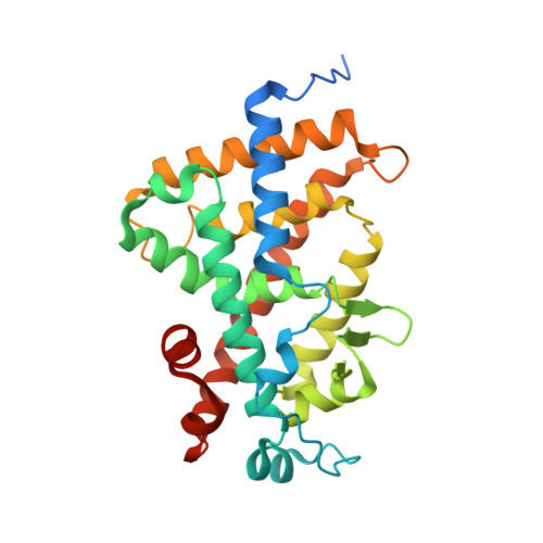
    An improved synthetic route to 1α,25-dihydroxyvitamin D(3) des-side chain analogues 2 a and 2 b with substituents at C18 is reported, along with their biological activity. These analogues display significant antiproliferative effects toward MCF-7 breast cancer cells and prodifferentiation activity toward SW480-ADH colon cancer cells; they are also characterized by a greatly decreased calcemic profile. The crystal structure of the human vitamin D receptor (hVDR) complexed to one of these analogues, 20(17→18)-abeo-1α,25-dihydroxy-22-homo-21-norvitamin D(3) (2 a) reveals that the side chain introduced at position C18 adopts the same orientation in the ligand binding pocket as the side chain of 1α,25-dihydroxyvitamin D(3).


  • Organizational Affiliation

    Laboratorium voor Experimentele Geneeskunde en Endocrinologie, Katholieke Universiteit Leuven, Leuven, Belgium.


Macromolecules
Find similar proteins by:  (by identity cutoff)  |  3D Structure
Entity ID: 1
MoleculeChains Sequence LengthOrganismDetailsImage
Vitamin D3 receptor280Homo sapiensMutation(s): 0 
Gene Names: VDRNR1I1
UniProt & NIH Common Fund Data Resources
Find proteins for P11473 (Homo sapiens)
Explore P11473 
Go to UniProtKB:  P11473
PHAROS:  P11473
GTEx:  ENSG00000111424 
Entity Groups  
Sequence Clusters30% Identity50% Identity70% Identity90% Identity95% Identity100% Identity
UniProt GroupP11473
Sequence Annotations
Expand
  • Reference Sequence
Small Molecules
Ligands 2 Unique
IDChains Name / Formula / InChI Key2D Diagram3D Interactions
ZYD
Query on ZYD

Download Ideal Coordinates CCD File 
B [auth A](1R,3S,5Z)-5-{(2E)-2-[(3aR,7aS)-7a-(7-hydroxy-7-methyloctyl)octahydro-4H-inden-4-ylidene]ethylidene}-4-methylidenecyclohexane-1,3-diol
C27 H44 O3
FJGYNKGWOKETAA-AXVRRUDCSA-N
SO4
Query on SO4

Download Ideal Coordinates CCD File 
C [auth A],
D [auth A]
SULFATE ION
O4 S
QAOWNCQODCNURD-UHFFFAOYSA-L
Experimental Data & Validation

Experimental Data

  • Method: X-RAY DIFFRACTION
  • Resolution: 1.70 Å
  • R-Value Free: 0.209 
  • R-Value Work: 0.189 
  • R-Value Observed: 0.189 
  • Space Group: P 21 21 21
Unit Cell:
Length ( Å )Angle ( ˚ )
a = 44.628α = 90
b = 51.342β = 90
c = 130.82γ = 90
Software Package:
Software NamePurpose
HKL-2000data collection
CNSrefinement
HKL-2000data reduction
HKL-2000data scaling
CNSphasing

Structure Validation

View Full Validation Report


Ligand Structure Quality Assessment 


Entry History 

Deposition Data

Revision History  (Full details and data files)

  • Version 1.0: 2011-08-31
    Type: Initial release
  • Version 1.1: 2017-07-26
    Changes: Advisory, Refinement description, Source and taxonomy
  • Version 1.2: 2024-02-21
    Changes: Advisory, Data collection, Database references, Derived calculations