3MOS

The structure of human Transketolase


Experimental Data Snapshot

  • Method: X-RAY DIFFRACTION
  • Resolution: 1.75 Å
  • R-Value Free: 0.206 
  • R-Value Work: 0.162 
  • R-Value Observed: 0.164 

wwPDB Validation   3D Report Full Report


Ligand Structure Quality Assessment 


This is version 1.2 of the entry. See complete history


Literature

The crystal structure of human transketolase and new insights into its mode of action.

Mitschke, L.Parthier, C.Schroder-Tittmann, K.Coy, J.Ludtke, S.Tittmann, K.

(2010) J Biol Chem 285: 31559-31570

  • DOI: https://doi.org/10.1074/jbc.M110.149955
  • Primary Citation of Related Structures:  
    3MOS

  • PubMed Abstract: 

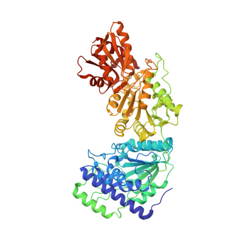
    The crystal structure of human transketolase (TKT), a thiamine diphosphate (ThDP) and Ca(2+)-dependent enzyme that catalyzes the interketol transfer between ketoses and aldoses as part of the pentose phosphate pathway, has been determined to 1.75 Å resolution. The recombinantly produced protein crystallized in space group C2 containing one monomer in the asymmetric unit. Two monomers form the homodimeric biological assembly with two identical active sites at the dimer interface. Although the protomer exhibits the typical three (α/β)-domain structure and topology reported for TKTs from other species, structural differences are observed for several loop regions and the linker that connects the PP and Pyr domain. The cofactor and substrate binding sites of human TKT bear high resemblance to those of other TKTs but also feature unique properties, including two lysines and a serine that interact with the β-phosphate of ThDP. Furthermore, Gln(189) spans over the thiazolium moiety of ThDP and replaces an isoleucine found in most non-mammalian TKTs. The side chain of Gln(428) forms a hydrogen bond with the 4'-amino group of ThDP and replaces a histidine that is invariant in all non-mammalian TKTs. All other amino acids involved in substrate binding and catalysis are strictly conserved. Besides a steady-state kinetic analysis, microscopic equilibria of the donor half-reaction were characterized by an NMR-based intermediate analysis. These studies reveal that formation of the central 1,2-dihydroxyethyl-ThDP carbanion-enamine intermediate is thermodynamically favored with increasing carbon chain length of the donor ketose substrate. Based on the structure of human transketolase and sequence alignments, putative functional properties of the related transketolase-like proteins TKTL1 and -2 are discussed in light of recent findings suggesting that TKTL1 plays a role in cancerogenesis.


  • Organizational Affiliation

    Institute of Biochemistry and Biotechnology, Martin-Luther-University Halle-Wittenberg, 06120 Halle, Germany.


Macromolecules
Find similar proteins by:  (by identity cutoff)  |  3D Structure
Entity ID: 1
MoleculeChains Sequence LengthOrganismDetailsImage
Transketolase616Homo sapiensMutation(s): 0 
Gene Names: TKT
EC: 2.2.1.1
UniProt & NIH Common Fund Data Resources
Find proteins for P29401 (Homo sapiens)
Explore P29401 
Go to UniProtKB:  P29401
PHAROS:  P29401
GTEx:  ENSG00000163931 
Entity Groups  
Sequence Clusters30% Identity50% Identity70% Identity90% Identity95% Identity100% Identity
UniProt GroupP29401
Sequence Annotations
Expand
  • Reference Sequence
Small Molecules
Ligands 4 Unique
IDChains Name / Formula / InChI Key2D Diagram3D Interactions
TPP
Query on TPP

Download Ideal Coordinates CCD File 
B [auth A]THIAMINE DIPHOSPHATE
C12 H19 N4 O7 P2 S
AYEKOFBPNLCAJY-UHFFFAOYSA-O
EDO
Query on EDO

Download Ideal Coordinates CCD File 
D [auth A]
E [auth A]
F [auth A]
G [auth A]
H [auth A]
D [auth A],
E [auth A],
F [auth A],
G [auth A],
H [auth A],
I [auth A],
J [auth A],
K [auth A],
L [auth A],
M [auth A]
1,2-ETHANEDIOL
C2 H6 O2
LYCAIKOWRPUZTN-UHFFFAOYSA-N
CA
Query on CA

Download Ideal Coordinates CCD File 
C [auth A]CALCIUM ION
Ca
BHPQYMZQTOCNFJ-UHFFFAOYSA-N
NA
Query on NA

Download Ideal Coordinates CCD File 
N [auth A]SODIUM ION
Na
FKNQFGJONOIPTF-UHFFFAOYSA-N
Experimental Data & Validation

Experimental Data

  • Method: X-RAY DIFFRACTION
  • Resolution: 1.75 Å
  • R-Value Free: 0.206 
  • R-Value Work: 0.162 
  • R-Value Observed: 0.164 
  • Space Group: C 1 2 1
Unit Cell:
Length ( Å )Angle ( ˚ )
a = 113.63α = 90
b = 85.33β = 125.69
c = 72.74γ = 90
Software Package:
Software NamePurpose
XSCALEdata scaling
PHASERphasing
REFMACrefinement
PDB_EXTRACTdata extraction
CrystalCleardata collection
XDSdata reduction

Structure Validation

View Full Validation Report


Ligand Structure Quality Assessment 


Entry History 

Deposition Data

Revision History  (Full details and data files)

  • Version 1.0: 2010-07-28
    Type: Initial release
  • Version 1.1: 2011-07-13
    Changes: Version format compliance
  • Version 1.2: 2023-09-06
    Changes: Data collection, Database references, Derived calculations, Refinement description