3MK0

Refinement of placental alkaline phosphatase complexed with nitrophenyl


Experimental Data Snapshot

  • Method: X-RAY DIFFRACTION
  • Resolution: 1.90 Å
  • R-Value Free: 0.190 
  • R-Value Work: 0.135 
  • R-Value Observed: 0.137 

wwPDB Validation   3D Report Full Report


This is version 2.0 of the entry. See complete history

Re-refinement Note

This entry reflects an alternative modeling of the original data in: 1zeb


Literature

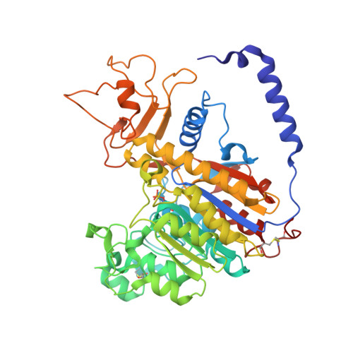
Refined structures of placental alkaline phosphatase show a consistent pattern of interactions at the peripheral site.

Stec, B.Cheltsov, A.Millan, J.L.

(2010) Acta Crystallogr Sect F Struct Biol Cryst Commun 66: 866-870

  • DOI: https://doi.org/10.1107/S1744309110019767
  • Primary Citation of Related Structures:  
    3MK0, 3MK1, 3MK2

  • PubMed Abstract: 

    In order to gain deeper insights into the functional sites of human placental alkaline phosphatase, the structures of the enzyme with the putative regulators L-Phe, pNPP and 5'-AMP [Llinas et al. (2005), J. Mol. Biol. 350, 441-451] were re-refined. Significant variations in ligand positioning and identity were found compared with the previous report. The multiple corrections to the model improved the phases and the electron-density maps, allowing the modeling of omitted side chains and multiple disordered residues. These improvements led to a change in the position of L-Phe at the peripheral binding site, which appeared to be reversed. The structure with pNPP contained only p-nitrophenol in three distinct sites, while the structure with 5'-AMP contained the p-nitrophenyl group in two of the sites instead of 5'-AMP. Comparison of the re-refined models shows a consistent pattern of interactions at the peripheral site.


  • Organizational Affiliation

    Sanford-Burnham Medical Research Institute, La Jolla, CA 92037, USA. bstec@burnham.org


Macromolecules
Find similar proteins by:  (by identity cutoff)  |  3D Structure
Entity ID: 1
MoleculeChains Sequence LengthOrganismDetailsImage
Alkaline phosphatase, placental type484Homo sapiensMutation(s): 0 
EC: 3.1.3.1
UniProt & NIH Common Fund Data Resources
Find proteins for P05187 (Homo sapiens)
Explore P05187 
Go to UniProtKB:  P05187
PHAROS:  P05187
GTEx:  ENSG00000163283 
Entity Groups  
Sequence Clusters30% Identity50% Identity70% Identity90% Identity95% Identity100% Identity
UniProt GroupP05187
Sequence Annotations
Expand
  • Reference Sequence
Oligosaccharides

Help

Entity ID: 2
MoleculeChains Length2D Diagram Glycosylation3D Interactions
2-acetamido-2-deoxy-beta-D-glucopyranose-(1-4)-2-acetamido-2-deoxy-beta-D-glucopyranose
B, C
2N-Glycosylation
Glycosylation Resources
GlyTouCan:  G42666HT
GlyCosmos:  G42666HT
GlyGen:  G42666HT
Small Molecules
Ligands 6 Unique
IDChains Name / Formula / InChI Key2D Diagram3D Interactions
NPO
Query on NPO

Download Ideal Coordinates CCD File 
H [auth A],
I [auth A]
P-NITROPHENOL
C6 H5 N O3
BTJIUGUIPKRLHP-UHFFFAOYSA-N
PO4
Query on PO4

Download Ideal Coordinates CCD File 
J [auth A]PHOSPHATE ION
O4 P
NBIIXXVUZAFLBC-UHFFFAOYSA-K
ZN
Query on ZN

Download Ideal Coordinates CCD File 
D [auth A],
E [auth A]
ZINC ION
Zn
PTFCDOFLOPIGGS-UHFFFAOYSA-N
ACT
Query on ACT

Download Ideal Coordinates CCD File 
K [auth A]ACETATE ION
C2 H3 O2
QTBSBXVTEAMEQO-UHFFFAOYSA-M
CA
Query on CA

Download Ideal Coordinates CCD File 
G [auth A]CALCIUM ION
Ca
BHPQYMZQTOCNFJ-UHFFFAOYSA-N
MG
Query on MG

Download Ideal Coordinates CCD File 
F [auth A]MAGNESIUM ION
Mg
JLVVSXFLKOJNIY-UHFFFAOYSA-N
Modified Residues  1 Unique
IDChains TypeFormula2D DiagramParent
SEP
Query on SEP
A
L-PEPTIDE LINKINGC3 H8 N O6 PSER
Experimental Data & Validation

Experimental Data

  • Method: X-RAY DIFFRACTION
  • Resolution: 1.90 Å
  • R-Value Free: 0.190 
  • R-Value Work: 0.135 
  • R-Value Observed: 0.137 
  • Space Group: C 2 2 21
Unit Cell:
Length ( Å )Angle ( ˚ )
a = 89.022α = 90
b = 113.894β = 90
c = 106.594γ = 90
Software Package:
Software NamePurpose
REFMACrefinement

Structure Validation

View Full Validation Report


Entry History 

Deposition Data

Revision History  (Full details and data files)

  • Version 1.0: 2011-01-19
    Type: Initial release
  • Version 1.1: 2011-07-13
    Changes: Version format compliance
  • Version 1.2: 2011-10-05
    Changes: Database references
  • Version 2.0: 2020-07-29
    Type: Remediation
    Reason: Carbohydrate remediation
    Changes: Atomic model, Data collection, Derived calculations, Structure summary