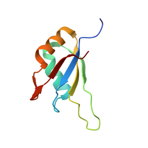
Molecular mechanism of MLL PHD3 and RNA recognition by the Cyp33 RRM domain.
Hom, R.A., Chang, P.Y., Roy, S., Musselman, C.A., Glass, K.C., Selezneva, A.I., Gozani, O., Ismagilov, R.F., Cleary, M.L., Kutateladze, T.G.(2010) J Mol Biol 400: 145-154
- PubMed: 20460131
- DOI: https://doi.org/10.1016/j.jmb.2010.04.067
- Primary Citation of Related Structures:
3MDF - PubMed Abstract:
The nuclear protein cyclophilin 33 (Cyp33) is a peptidyl-prolyl cis-trans isomerase that catalyzes cis-trans isomerization of the peptide bond preceding a proline and promotes folding and conformational changes in folded and unfolded proteins. The N-terminal RNA-recognition motif (RRM) domain of Cyp33 has been found to associate with the third plant homeodomain (PHD3) finger of the mixed lineage leukemia (MLL) proto-oncoprotein and a poly(A) RNA sequence. Here, we report a 1.9 A resolution crystal structure of the RRM domain of Cyp33 and describe the molecular mechanism of PHD3 and RNA recognition. The Cyp33 RRM domain folds into a five-stranded antiparallel beta-sheet and two alpha-helices. The RRM domain, but not the catalytic module of Cyp33, binds strongly to PHD3, exhibiting a 2 muM affinity as measured by isothermal titration calorimetry. NMR chemical shift perturbation (CSP) analysis and dynamics data reveal that the beta strands and the beta2-beta3 loop of the RRM domain are involved in the interaction with PHD3. Mutations in the PHD3-binding site or deletions in the beta2-beta3 loop lead to a significantly reduced affinity or abrogation of the interaction. The RNA-binding pocket of the Cyp33 RRM domain, mapped on the basis of NMR CSP and mutagenesis, partially overlaps with the PHD3-binding site, and RNA association is abolished in the presence of MLL PHD3. Full-length Cyp33 acts as a negative regulator of MLL-induced transcription and reduces the expression levels of MLL target genes MEIS1 and HOXA9. Together, these in vitro and in vivo data provide insight into the multiple functions of Cyp33 RRM and suggest a Cyp33-dependent mechanism for regulating the transcriptional activity of MLL.
Organizational Affiliation:
Department of Pharmacology, University of Colorado Denver School of Medicine, 12801 East 17th Avenue, Aurora, CO 80045, USA.