3KQ4

Structure of Intrinsic Factor-Cobalamin bound to its receptor Cubilin


Experimental Data Snapshot

  • Method: X-RAY DIFFRACTION
  • Resolution: 3.30 Å
  • R-Value Free: 0.242 
  • R-Value Work: 0.211 
  • R-Value Observed: 0.211 

wwPDB Validation   3D Report Full Report


Ligand Structure Quality Assessment 


This is version 2.0 of the entry. See complete history


Literature

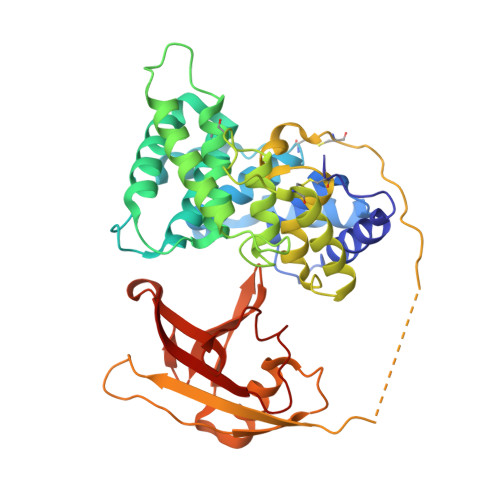
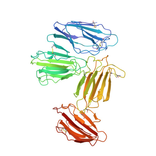
Structural basis for receptor recognition of vitamin-B(12)-intrinsic factor complexes.

Andersen, C.B.Madsen, M.Storm, T.Moestrup, S.K.Andersen, G.R.

(2010) Nature 464: 445-448

  • DOI: https://doi.org/10.1038/nature08874
  • Primary Citation of Related Structures:  
    3KQ4

  • PubMed Abstract: 

    Cobalamin (Cbl, vitamin B(12)) is a bacterial organic compound and an essential coenzyme in mammals, which take it up from the diet. This occurs by the combined action of the gastric intrinsic factor (IF) and the ileal endocytic cubam receptor formed by the 460-kilodalton (kDa) protein cubilin and the 45-kDa transmembrane protein amnionless. Loss of function of any of these proteins ultimately leads to Cbl deficiency in man. Here we present the crystal structure of the complex between IF-Cbl and the cubilin IF-Cbl-binding-region (CUB(5-8)) determined at 3.3 A resolution. The structure provides insight into how several CUB (for 'complement C1r/C1s, Uegf, Bmp1') domains collectively function as modular ligand-binding regions, and how two distant CUB domains embrace the Cbl molecule by binding the two IF domains in a Ca(2+)-dependent manner. This dual-point model provides a probable explanation of how Cbl indirectly induces ligand-receptor coupling. Finally, the comparison of Ca(2+)-binding CUB domains and the low-density lipoprotein (LDL) receptor-type A modules suggests that the electrostatic pairing of a basic ligand arginine/lysine residue with Ca(2+)-coordinating acidic aspartates/glutamates is a common theme of Ca(2+)-dependent ligand-receptor interactions.


  • Organizational Affiliation

    Department of Medical Biochemistry, Aarhus University, 8000 Aarhus C, Denmark.


Macromolecules
Find similar proteins by:  (by identity cutoff)  |  3D Structure
Entity ID: 1
MoleculeChains Sequence LengthOrganismDetailsImage
Gastric intrinsic factor
A, C, E
393Homo sapiensMutation(s): 0 
Gene Names: GIFIFMH
UniProt & NIH Common Fund Data Resources
Find proteins for P27352 (Homo sapiens)
Explore P27352 
Go to UniProtKB:  P27352
PHAROS:  P27352
GTEx:  ENSG00000134812 
Entity Groups  
Sequence Clusters30% Identity50% Identity70% Identity90% Identity95% Identity100% Identity
UniProt GroupP27352
Sequence Annotations
Expand
  • Reference Sequence
Find similar proteins by:  (by identity cutoff)  |  3D Structure
Entity ID: 2
MoleculeChains Sequence LengthOrganismDetailsImage
Cubilin
B, D, F
457Homo sapiensMutation(s): 0 
Gene Names: CUBNIFCR
UniProt & NIH Common Fund Data Resources
Find proteins for O60494 (Homo sapiens)
Explore O60494 
Go to UniProtKB:  O60494
PHAROS:  O60494
GTEx:  ENSG00000107611 
Entity Groups  
Sequence Clusters30% Identity50% Identity70% Identity90% Identity95% Identity100% Identity
UniProt GroupO60494
Sequence Annotations
Expand
  • Reference Sequence
Oligosaccharides

Help

Entity ID: 3
MoleculeChains Length2D Diagram Glycosylation3D Interactions
alpha-D-mannopyranose-(1-3)-[alpha-D-mannopyranose-(1-6)]beta-D-mannopyranose-(1-4)-2-acetamido-2-deoxy-beta-D-glucopyranose-(1-4)-2-acetamido-2-deoxy-beta-D-glucopyranose
G, H, J, K, M
G, H, J, K, M, N
5N-Glycosylation
Glycosylation Resources
GlyTouCan:  G22768VO
GlyCosmos:  G22768VO
GlyGen:  G22768VO
Entity ID: 4
MoleculeChains Length2D Diagram Glycosylation3D Interactions
2-acetamido-2-deoxy-beta-D-glucopyranose-(1-4)-2-acetamido-2-deoxy-beta-D-glucopyranose
I, L, O
2N-Glycosylation
Glycosylation Resources
GlyTouCan:  G42666HT
GlyCosmos:  G42666HT
GlyGen:  G42666HT
Small Molecules
Ligands 3 Unique
IDChains Name / Formula / InChI Key2D Diagram3D Interactions
B12
Query on B12

Download Ideal Coordinates CCD File 
CA [auth C],
OA [auth E],
Q [auth A]
COBALAMIN
C62 H89 Co N13 O14 P
LKVIQTCSMMVGFU-DWSMJLPVSA-N
NAG
Query on NAG

Download Ideal Coordinates CCD File 
BA [auth C]
DA [auth D]
EA [auth D]
FA [auth D]
GA [auth D]
BA [auth C],
DA [auth D],
EA [auth D],
FA [auth D],
GA [auth D],
HA [auth D],
IA [auth D],
NA [auth E],
P [auth A],
PA [auth F],
QA [auth F],
R [auth B],
RA [auth F],
S [auth B],
SA [auth F],
T [auth B],
TA [auth F],
U [auth B],
UA [auth F],
V [auth B],
W [auth B]
2-acetamido-2-deoxy-beta-D-glucopyranose
C8 H15 N O6
OVRNDRQMDRJTHS-FMDGEEDCSA-N
CA
Query on CA

Download Ideal Coordinates CCD File 
AA [auth B]
JA [auth D]
KA [auth D]
LA [auth D]
MA [auth D]
AA [auth B],
JA [auth D],
KA [auth D],
LA [auth D],
MA [auth D],
VA [auth F],
WA [auth F],
X [auth B],
XA [auth F],
Y [auth B],
YA [auth F],
Z [auth B]
CALCIUM ION
Ca
BHPQYMZQTOCNFJ-UHFFFAOYSA-N
Experimental Data & Validation

Experimental Data

  • Method: X-RAY DIFFRACTION
  • Resolution: 3.30 Å
  • R-Value Free: 0.242 
  • R-Value Work: 0.211 
  • R-Value Observed: 0.211 
  • Space Group: C 2 2 21
Unit Cell:
Length ( Å )Angle ( ˚ )
a = 117.684α = 90
b = 204.179β = 90
c = 410.023γ = 90
Software Package:
Software NamePurpose
PHENIXrefinement

Structure Validation

View Full Validation Report


Ligand Structure Quality Assessment 


Entry History 

Deposition Data

Revision History  (Full details and data files)

  • Version 1.0: 2010-03-09
    Type: Initial release
  • Version 1.1: 2011-07-13
    Changes: Version format compliance
  • Version 1.2: 2012-10-24
    Changes: Non-polymer description
  • Version 1.3: 2018-03-07
    Changes: Advisory, Data collection
  • Version 2.0: 2020-07-29
    Type: Remediation
    Reason: Carbohydrate remediation
    Changes: Advisory, Atomic model, Data collection, Derived calculations, Structure summary