3KC0

Crystal structure of human liver FBPase in complex with tricyclic inhibitor 10b


Experimental Data Snapshot

  • Method: X-RAY DIFFRACTION
  • Resolution: 2.80 Å
  • R-Value Free: 0.254 
  • R-Value Work: 0.205 

wwPDB Validation   3D Report Full Report


Ligand Structure Quality Assessment 


This is version 1.3 of the entry. See complete history


Literature

Structure-based drug design of tricyclic 8H-indeno[1,2-d][1,3]thiazoles as potent FBPase inhibitors.

Tsukada, T.Takahashi, M.Takemoto, T.Kanno, O.Yamane, T.Kawamura, S.Nishi, T.

(2010) Bioorg Med Chem Lett 20: 1004-1007

  • DOI: https://doi.org/10.1016/j.bmcl.2009.12.056
  • Primary Citation of Related Structures:  
    3KBZ, 3KC0, 3KC1

  • PubMed Abstract: 

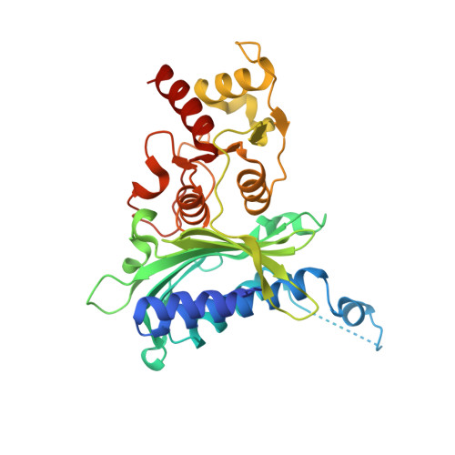
    With the goal of improving metabolic stability and further enhancing FBPase inhibitory activity, a series of tricyclic 8H-indeno[1,2-d][1,3]thiazoles was designed and synthesized with the aid of structure-based drug design. Extensive SAR studies led to the discovery of 19a with an IC(50) value of 1nM against human FBPase. X-ray crystallographic studies revealed that high affinity of 19a was due to the hydrophobic interaction arising from better shape complementarity and to the hydrogen bonding network involving the side chain on the tricyclic scaffold.


  • Organizational Affiliation

    Medicinal Chemistry Research Laboratories I, Daiichi Sankyo Co, Ltd, 1-2-58 Hiromachi, Shinagawa-ku, Tokyo 140-8710, Japan.


Macromolecules
Find similar proteins by:  (by identity cutoff)  |  3D Structure
Entity ID: 1
MoleculeChains Sequence LengthOrganismDetailsImage
Fructose-1,6-bisphosphatase 1
A, B, C, D
337Homo sapiensMutation(s): 0 
EC: 3.1.3.11
UniProt & NIH Common Fund Data Resources
Find proteins for P09467 (Homo sapiens)
Explore P09467 
Go to UniProtKB:  P09467
PHAROS:  P09467
GTEx:  ENSG00000165140 
Entity Groups  
Sequence Clusters30% Identity50% Identity70% Identity90% Identity95% Identity100% Identity
UniProt GroupP09467
Sequence Annotations
Expand
  • Reference Sequence
Small Molecules
Binding Affinity Annotations 
IDSourceBinding Affinity
2T5 PDBBind:  3KC0 IC50: 12 (nM) from 1 assay(s)
BindingDB:  3KC0 IC50: 12 (nM) from 1 assay(s)
Experimental Data & Validation

Experimental Data

  • Method: X-RAY DIFFRACTION
  • Resolution: 2.80 Å
  • R-Value Free: 0.254 
  • R-Value Work: 0.205 
  • Space Group: P 21 21 21
Unit Cell:
Length ( Å )Angle ( ˚ )
a = 67.246α = 90
b = 82.82β = 90
c = 276.631γ = 90
Software Package:
Software NamePurpose
DENZOdata reduction
SCALEPACKdata scaling
CNSrefinement
PDB_EXTRACTdata extraction
SERGUIdata collection
CNXphasing
CNXrefinement

Structure Validation

View Full Validation Report


Ligand Structure Quality Assessment 


Entry History 

Deposition Data

Revision History  (Full details and data files)

  • Version 1.0: 2010-02-02
    Type: Initial release
  • Version 1.1: 2011-07-13
    Changes: Version format compliance
  • Version 1.2: 2017-11-01
    Changes: Refinement description
  • Version 1.3: 2023-11-01
    Changes: Data collection, Database references, Derived calculations, Refinement description