3KBT

Crystal structure of the ankyrin binding domain of human erythroid beta spectrin (repeats 13-15) in complex with the spectrin binding domain of human erythroid ankyrin (ZU5-ANK)


Experimental Data Snapshot

  • Method: X-RAY DIFFRACTION
  • Resolution: 2.75 Å
  • R-Value Free: 0.292 
  • R-Value Work: 0.245 
  • R-Value Observed: 0.247 

wwPDB Validation   3D Report Full Report


This is version 1.3 of the entry. See complete history


Literature

Structural basis for spectrin recognition by ankyrin.

Ipsaro, J.J.Mondragon, A.

(2010) Blood 115: 4093-4101

  • DOI: https://doi.org/10.1182/blood-2009-11-255604
  • Primary Citation of Related Structures:  
    3KBT, 3KBU

  • PubMed Abstract: 

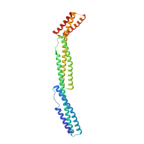
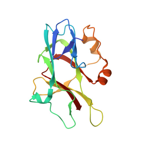
    Maintenance of membrane integrity and organization in the metazoan cell is accomplished through intracellular tethering of membrane proteins to an extensive, flexible protein network. Spectrin, the principal component of this network, is anchored to membrane proteins through the adaptor protein ankyrin. To elucidate the atomic basis for this interaction, we determined a crystal structure of human betaI-spectrin repeats 13 to 15 in complex with the ZU5-ANK domain of human ankyrin R. The structure reveals the role of repeats 14 to 15 in binding, the electrostatic and hydrophobic contributions along the interface, and the necessity for a particular orientation of the spectrin repeats. Using structural and biochemical data as a guide, we characterized the individual proteins and their interactions by binding and thermal stability analyses. In addition to validating the structural model, these data provide insight into the nature of some mutations associated with cell morphology defects, including those found in human diseases such as hereditary spherocytosis and elliptocytosis. Finally, analysis of the ZU5 domain suggests it is a versatile protein-protein interaction module with distinct interaction surfaces. The structure represents not only the first of a spectrin fragment in complex with its binding partner, but also that of an intermolecular complex involving a ZU5 domain.


  • Organizational Affiliation

    Department of Biochemistry, Molecular Biology, and Cell Biology, Northwestern University, Evanston, IL 60208, USA.


Macromolecules
Find similar proteins by:  (by identity cutoff)  |  3D Structure
Entity ID: 1
MoleculeChains Sequence LengthOrganismDetailsImage
Spectrin beta chain, erythrocyte
A, B
326Homo sapiensMutation(s): 0 
Gene Names: SPTBSPTB1
UniProt & NIH Common Fund Data Resources
Find proteins for P11277 (Homo sapiens)
Explore P11277 
Go to UniProtKB:  P11277
PHAROS:  P11277
GTEx:  ENSG00000070182 
Entity Groups  
Sequence Clusters30% Identity50% Identity70% Identity90% Identity95% Identity100% Identity
UniProt GroupP11277
Sequence Annotations
Expand
  • Reference Sequence
Find similar proteins by:  (by identity cutoff)  |  3D Structure
Entity ID: 2
MoleculeChains Sequence LengthOrganismDetailsImage
Ankyrin-1
C, D
161Homo sapiensMutation(s): 0 
Gene Names: ANKANK1
UniProt & NIH Common Fund Data Resources
Find proteins for P16157 (Homo sapiens)
Explore P16157 
Go to UniProtKB:  P16157
PHAROS:  P16157
GTEx:  ENSG00000029534 
Entity Groups  
Sequence Clusters30% Identity50% Identity70% Identity90% Identity95% Identity100% Identity
UniProt GroupP16157
Sequence Annotations
Expand
  • Reference Sequence
Experimental Data & Validation

Experimental Data

  • Method: X-RAY DIFFRACTION
  • Resolution: 2.75 Å
  • R-Value Free: 0.292 
  • R-Value Work: 0.245 
  • R-Value Observed: 0.247 
  • Space Group: P 21 21 21
Unit Cell:
Length ( Å )Angle ( ˚ )
a = 90.45α = 90
b = 95.66β = 90
c = 137.93γ = 90
Software Package:
Software NamePurpose
MAR345data collection
SHARPphasing
REFMACrefinement
XDSdata reduction
SCALAdata scaling

Structure Validation

View Full Validation Report


Entry History 

Deposition Data

Revision History  (Full details and data files)

  • Version 1.0: 2010-02-02
    Type: Initial release
  • Version 1.1: 2011-07-13
    Changes: Advisory, Refinement description, Version format compliance
  • Version 1.2: 2017-11-01
    Changes: Refinement description
  • Version 1.3: 2024-02-21
    Changes: Data collection, Database references