


3HV3
Human p38 MAP Kinase in Complex with RL49
- PDB DOI: https://doi.org/10.2210/pdb3HV3/pdb
- Classification: TRANSFERASE
- Organism(s): Homo sapiens
- Expression System: Escherichia coli
- Mutation(s): Yes
- Deposited: 2009-06-15 Released: 2009-11-17
Experimental Data Snapshot
- Method: X-RAY DIFFRACTION
- Resolution: 2.00 Å
- R-Value Free: 0.300
- R-Value Work: 0.230
- R-Value Observed: 0.233

This is version 1.4 of the entry. See complete history.

Macromolecules
Find similar proteins by:
(by identity cutoff) | 3D Structure
Entity ID: 1 | |||||
|---|---|---|---|---|---|
| Molecule | Chains | Sequence Length | Organism | Details | Image |
| Mitogen-activated protein kinase 14 | 360 | Homo sapiens | Mutation(s): 4 Gene Names: MAPK14, CSBP, CSBP1, CSBP2, CSPB1, MXI2 EC: 2.7.11.24 | 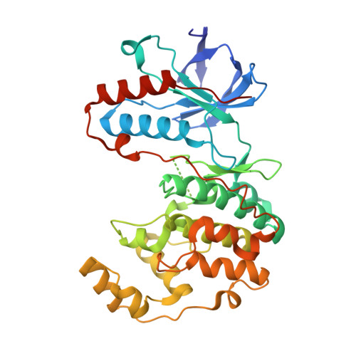 | |
UniProt & NIH Common Fund Data Resources | |||||
Find proteins for Q16539 (Homo sapiens) Explore Q16539 Go to UniProtKB: Q16539 | |||||
PHAROS: Q16539 GTEx: ENSG00000112062 | |||||
Entity Groups | |||||
| Sequence Clusters | 30% Identity50% Identity70% Identity90% Identity95% Identity100% Identity | ||||
| UniProt Group | Q16539 | ||||
Sequence AnnotationsExpand | |||||
| |||||
Small Molecules
| Ligands 3 Unique | |||||
|---|---|---|---|---|---|
| ID | Chains | Name / Formula / InChI Key | 2D Diagram | 3D Interactions | |
| R49 Query on R49 | B [auth A] | 1-{4-[(6-aminoquinolin-4-yl)amino]phenyl}-3-[3-tert-butyl-1-(3-methylphenyl)-1H-pyrazol-5-yl]urea C30 H31 N7 O MKHUGLZLKVCOGF-UHFFFAOYSA-N |  | ||
| BOG Query on BOG | C [auth A] | octyl beta-D-glucopyranoside C14 H28 O6 HEGSGKPQLMEBJL-RKQHYHRCSA-N |  | ||
| GOL Query on GOL | D [auth A] | GLYCEROL C3 H8 O3 PEDCQBHIVMGVHV-UHFFFAOYSA-N |  | ||
Experimental Data & Validation
Experimental Data
- Method: X-RAY DIFFRACTION
- Resolution: 2.00 Å
- R-Value Free: 0.300
- R-Value Work: 0.230
- R-Value Observed: 0.233
- Space Group: P 21 21 21
Unit Cell:
| Length ( Å ) | Angle ( ˚ ) |
|---|---|
| a = 66.51 | α = 90 |
| b = 74.52 | β = 90 |
| c = 76.98 | γ = 90 |
| Software Name | Purpose |
|---|---|
| XSCALE | data scaling |
| PHASER | phasing |
| REFMAC | refinement |
| PDB_EXTRACT | data extraction |
| XDS | data scaling |
Entry History
Deposition Data
- Released Date: 2009-11-17 Deposition Author(s): Gruetter, C., Simard, J.R., Getlik, M., Rauh, D.
Revision History (Full details and data files)
- Version 1.0: 2009-11-17
Type: Initial release - Version 1.1: 2011-07-13
Changes: Non-polymer description, Version format compliance - Version 1.2: 2020-07-29
Type: Remediation
Reason: Carbohydrate remediation
Changes: Data collection, Database references, Derived calculations, Structure summary - Version 1.3: 2021-10-13
Changes: Database references, Structure summary - Version 1.4: 2023-09-06
Changes: Data collection, Refinement description
