Skeletal dysplasias due to filamin A mutations result from a gain-of-function mechanism distinct from allelic neurological disorders
Clark, A.R., Sawyer, G.M., Robertson, S.P., Sutherland-Smith, A.J.(2009) Hum Mol Genet 18: 4791-4800
- PubMed: 19773341
- DOI: https://doi.org/10.1093/hmg/ddp442
- Primary Citation of Related Structures:
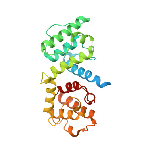
3HOC, 3HOP, 3HOR - PubMed Abstract:
Filamin A (FLNA) crosslinks F-actin and binds proteins consistent with roles integrating cell signalling and the cytoskeleton. FLNA missense mutations are associated with the otopalatodigital syndrome (OPD) spectrum of skeletal disorders, clustering in discrete domains. One cluster is found in the second calponin homology domain of the FLNA actin-binding domain (ABD), implicating this region as essential for mediating correct function. Here we show that OPD (FLNA E254K) fibroblast lysates have equivalent concentrations of FLNA compared with controls and that recombinant FLNA E254K ABD has increased in vitro F-actin binding (K(d) 13 microm) compared with wild type (WT; K(d) 48 microm). These observations are consistent with a gain-of-function mechanism for OPD. We have determined the crystal structures of the WT and E254K FLNA ABDs at 2.3 A resolution, revealing that they adopt similar closed conformations. The E254K mutation removes a conserved salt bridge but does not disrupt the ABD structure. The solution structures are also equivalent as determined by circular dichroism spectroscopy, but differential scanning fluorimetry denaturation showed reduced stability (decreased T(m) of 5.6 degrees C) for E254K relative to WT. Ex vivo characterization of E254K OPD patient fibroblasts revealed they have similar motility and adhesion as control cells, implying that many core functions mediated by FLNA are unaffected, consistent with OPD only affecting specific tissues despite FLNA being widely expressed. These data provide the first biochemical evidence for a gain-of-function mechanism for the OPD disorders, and mechanistically distinguishes them from the loss-of-function phenotypes that manifest as disorders of neuronal migration.
Organizational Affiliation:
Institute of Molecular BioSciences, Massey University, Palmerston North, New Zealand.