3EZP

Crystal Structure Analysis of human HDAC8 D101N variant


Experimental Data Snapshot

  • Method: X-RAY DIFFRACTION
  • Resolution: 2.65 Å
  • R-Value Free: 0.262 
  • R-Value Work: 0.225 
  • R-Value Observed: 0.225 

wwPDB Validation   3D Report Full Report


Ligand Structure Quality Assessment 


This is version 1.4 of the entry. See complete history


Literature

Structural studies of human histone deacetylase 8 and its site-specific variants complexed with substrate and inhibitors.

Dowling, D.P.Gantt, S.L.Gattis, S.G.Fierke, C.A.Christianson, D.W.

(2008) Biochemistry 47: 13554-13563

  • DOI: https://doi.org/10.1021/bi801610c
  • Primary Citation of Related Structures:  
    3EW8, 3EWF, 3EZP, 3EZT, 3F06, 3F07, 3F0R

  • PubMed Abstract: 

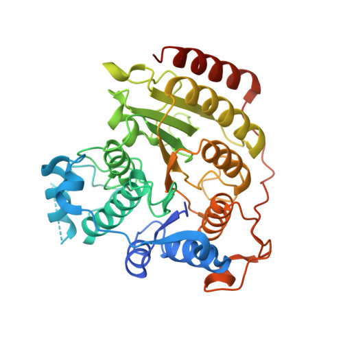
    Metal-dependent histone deacetylases (HDACs) require Zn(2+) or Fe(2+) to regulate the acetylation of lysine residues in histones and other proteins in eukaryotic cells. Isozyme HDAC8 is perhaps the archetypical member of the class I HDAC family and serves as a paradigm for studying structure-function relationships. Here, we report the structures of HDAC8 complexes with trichostatin A and 3-(1-methyl-4-phenylacetyl-1H-2-pyrrolyl)-N-hydroxy-2-propenamide (APHA) in a new crystal form. The structure of the APHA complex reveals that the hydroxamate CO group accepts a hydrogen bond from Y306 but does not coordinate to Zn(2+) with favorable geometry, perhaps due to the constraints of its extended pi system. Additionally, since APHA binds to only two of the three protein molecules in the asymmetric unit of this complex, the structure of the third monomer represents the first structure of HDAC8 in the unliganded state. Comparison of unliganded and liganded structures illustrates ligand-induced conformational changes in the L2 loop that likely accompany substrate binding and catalysis. Furthermore, these structures, along with those of the D101N, D101E, D101A, and D101L variants, support the proposal that D101 is critical for the function of the L2 loop. However, amino acid substitutions for D101 can also trigger conformational changes of Y111 and W141 that perturb the substrate binding site. Finally, the structure of H143A HDAC8 complexed with an intact acetylated tetrapeptide substrate molecule confirms the importance of D101 for substrate binding and reveals how Y306 and the active site zinc ion together bind and activate the scissile amide linkage of acetyllysine.


  • Organizational Affiliation

    Roy and Diana Vagelos Laboratories, Department of Chemistry, University of Pennsylvania, Philadelphia, Pennsylvania 19104-6323, USA.


Macromolecules
Find similar proteins by:  (by identity cutoff)  |  3D Structure
Entity ID: 1
MoleculeChains Sequence LengthOrganismDetailsImage
Histone deacetylase 8
A, B
388Homo sapiensMutation(s): 1 
Gene Names: CDA07HDAC8HDACL1
EC: 3.5.1.98
UniProt & NIH Common Fund Data Resources
Find proteins for Q9BY41 (Homo sapiens)
Explore Q9BY41 
Go to UniProtKB:  Q9BY41
PHAROS:  Q9BY41
GTEx:  ENSG00000147099 
Entity Groups  
Sequence Clusters30% Identity50% Identity70% Identity90% Identity95% Identity100% Identity
UniProt GroupQ9BY41
Sequence Annotations
Expand
  • Reference Sequence
Small Molecules
Ligands 4 Unique
IDChains Name / Formula / InChI Key2D Diagram3D Interactions
B3N
Query on B3N

Download Ideal Coordinates CCD File 
F [auth A],
K [auth B]
4-(dimethylamino)-N-[7-(hydroxyamino)-7-oxoheptyl]benzamide
C16 H25 N3 O3
MXWDSZWTBOCWBK-UHFFFAOYSA-N
BME
Query on BME

Download Ideal Coordinates CCD File 
G [auth A],
L [auth B]
BETA-MERCAPTOETHANOL
C2 H6 O S
DGVVWUTYPXICAM-UHFFFAOYSA-N
ZN
Query on ZN

Download Ideal Coordinates CCD File 
C [auth A],
H [auth B]
ZINC ION
Zn
PTFCDOFLOPIGGS-UHFFFAOYSA-N
K
Query on K

Download Ideal Coordinates CCD File 
D [auth A],
E [auth A],
I [auth B],
J [auth B]
POTASSIUM ION
K
NPYPAHLBTDXSSS-UHFFFAOYSA-N
Experimental Data & Validation

Experimental Data

  • Method: X-RAY DIFFRACTION
  • Resolution: 2.65 Å
  • R-Value Free: 0.262 
  • R-Value Work: 0.225 
  • R-Value Observed: 0.225 
  • Space Group: P 1 21 1
Unit Cell:
Length ( Å )Angle ( ˚ )
a = 55.31α = 90
b = 85.874β = 93.6
c = 94.642γ = 90
Software Package:
Software NamePurpose
CNSrefinement
CBASSdata collection
HKL-2000data reduction
HKL-2000data scaling
PHASERphasing

Structure Validation

View Full Validation Report


Ligand Structure Quality Assessment 


Entry History 

Deposition Data

Revision History  (Full details and data files)

  • Version 1.0: 2008-12-30
    Type: Initial release
  • Version 1.1: 2011-07-13
    Changes: Non-polymer description, Version format compliance
  • Version 1.2: 2020-12-30
    Changes: Database references, Derived calculations, Structure summary
  • Version 1.3: 2021-10-20
    Changes: Database references
  • Version 1.4: 2023-09-06
    Changes: Data collection, Refinement description