3B7T

[E296Q]LTA4H in complex with Arg-Ala-Arg substrate


Experimental Data Snapshot

  • Method: X-RAY DIFFRACTION
  • Resolution: 2.30 Å
  • R-Value Free: 0.273 
  • R-Value Work: 0.206 

wwPDB Validation   3D Report Full Report


This is version 1.5 of the entry. See complete history


Literature

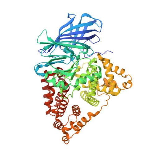
Structure-based dissection of the active site chemistry of leukotriene a4 hydrolase: implications for m1 aminopeptidases and inhibitor design.

Tholander, F.Muroya, A.Roques, B.P.Fournie-Zaluski, M.C.Thunnissen, M.M.Haeggstrom, J.Z.

(2008) Chem Biol 15: 920-929

  • DOI: https://doi.org/10.1016/j.chembiol.2008.07.018
  • Primary Citation of Related Structures:  
    2R59, 3B7R, 3B7S, 3B7T, 3B7U

  • PubMed Abstract: 

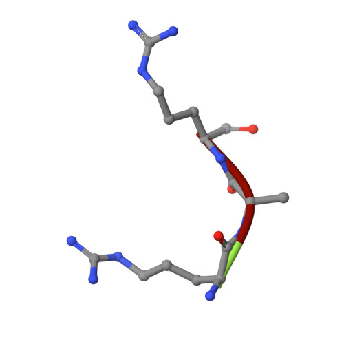
    M1 aminopeptidases comprise a large family of biologically important zinc enzymes. We show that peptide turnover by the M1 prototype, leukotriene A4 hydrolase/aminopeptidase, involves a shift in substrate position associated with exchange of zinc coordinating groups, while maintaining the overall coordination geometry. The transition state is stabilized by residues conserved among M1 members and in the final reaction step, Glu-296 of the canonical zinc binding HEXXH motif shuffles a proton from the hydrolytic water to the leaving group. Tripeptide substrates bind along the conserved GXMEN motif, precisely occupying the distance between Glu-271 and Arg-563, whereas the Arg specificity is governed by a narrow S1 pocket capped with Asp-375. Our data provide detailed insights to the active site chemistry of M1 aminopeptidases and will aid in the development of novel enzyme inhibitors.


  • Organizational Affiliation

    Department of Medical Biochemistry and Biophysics, Division of Chemistry II, Karolinska Institute, Stockholm, Sweden.


Macromolecules
Find similar proteins by:  (by identity cutoff)  |  3D Structure
Entity ID: 1
MoleculeChains Sequence LengthOrganismDetailsImage
Leukotriene A-4 hydrolase616Homo sapiensMutation(s): 1 
Gene Names: LTA4HLTA4
EC: 3.3.2.6 (PDB Primary Data), 3.4.11.4 (PDB Primary Data)
UniProt & NIH Common Fund Data Resources
Find proteins for P09960 (Homo sapiens)
Explore P09960 
Go to UniProtKB:  P09960
PHAROS:  P09960
GTEx:  ENSG00000111144 
Entity Groups  
Sequence Clusters30% Identity50% Identity70% Identity90% Identity95% Identity100% Identity
UniProt GroupP09960
Sequence Annotations
Expand
  • Reference Sequence

Find similar proteins by:  Sequence   |   3D Structure  

Entity ID: 2
MoleculeChains Sequence LengthOrganismDetailsImage
RAR peptide3N/AMutation(s): 0 
Sequence Annotations
Expand
  • Reference Sequence
Experimental Data & Validation

Experimental Data

  • Method: X-RAY DIFFRACTION
  • Resolution: 2.30 Å
  • R-Value Free: 0.273 
  • R-Value Work: 0.206 
  • Space Group: P 21 21 21
Unit Cell:
Length ( Å )Angle ( ˚ )
a = 78.49α = 90
b = 87.785β = 90
c = 99.916γ = 90
Software Package:
Software NamePurpose
XSCALEdata scaling
CNSrefinement
PDB_EXTRACTdata extraction
MAR345dtbdata collection
XDSdata reduction
XDSdata scaling
XFITdata reduction

Structure Validation

View Full Validation Report


Entry History 

Deposition Data

Revision History  (Full details and data files)

  • Version 1.0: 2008-09-16
    Type: Initial release
  • Version 1.1: 2011-07-13
    Changes: Version format compliance
  • Version 1.2: 2018-03-07
    Changes: Data collection
  • Version 1.3: 2019-11-20
    Changes: Database references, Derived calculations
  • Version 1.4: 2021-10-20
    Changes: Database references, Derived calculations, Source and taxonomy, Structure summary
  • Version 1.5: 2023-08-30
    Changes: Data collection, Refinement description