2Z6O

Crystal Structure of the Ufc1, Ufm1 conjugating enzyme 1


Experimental Data Snapshot

  • Method: X-RAY DIFFRACTION
  • Resolution: 1.60 Å
  • R-Value Free: 0.221 
  • R-Value Work: 0.194 
  • R-Value Observed: 0.196 

wwPDB Validation   3D Report Full Report


This is version 1.2 of the entry. See complete history


Literature

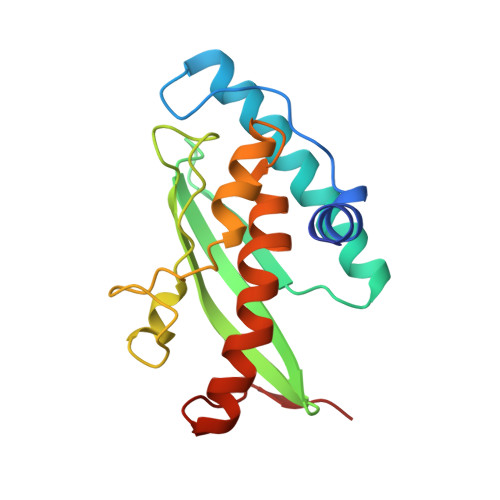
Crystal structure of Ufc1, the Ufm1-conjugating enzyme

Mizushima, T.Tatsumi, K.Ozaki, Y.Kawakami, T.Suzuki, A.Ogasahara, K.Komatsu, M.Kominami, E.Tanaka, K.Yamane, T.

(2007) Biochem Biophys Res Commun 362: 1079-1084

  • DOI: https://doi.org/10.1016/j.bbrc.2007.08.129
  • Primary Citation of Related Structures:  
    2Z6O, 2Z6P

  • PubMed Abstract: 

    Ubiquitin and ubiquitin-like protein-conjugating enzymes play central roles in posttranslational modification processes. The ubiquitin-fold modifier 1 (Ufm1), one of a variety of ubiquitin-like modifiers, is covalently attached to target proteins via Uba5 and Ufm1-conjugating enzyme 1 (Ufc1), which are analogous to the E1 and E2 ubiquitylation enzymes. As Ufm1-related proteins are conserved in metazoa and plants, the Ufm1 system likely plays important roles in various multicellular organisms. Herein, we report the X-ray structure of human Ufc1 determined at 1.6 A resolution. The Ufc1 structure comprises a canonical E2 domain and an additional N-terminal domain. The Uba5 binding site on Ufc1 was assigned by structural comparison of Ufc1 and Ubc12 and related mutational analyses. In addition, we show that the N-terminal unique domain of Ufc1 contributes to thermal stability.


  • Organizational Affiliation

    Department of Biotechnology, Graduate School of Engineering, Nagoya University, Chikusa-ku, Nagoya 464-8603, Japan.


Macromolecules
Find similar proteins by:  (by identity cutoff)  |  3D Structure
Entity ID: 1
MoleculeChains Sequence LengthOrganismDetailsImage
Ufm1-conjugating enzyme 1172Homo sapiensMutation(s): 0 
Gene Names: UFC1
EC: 6.3.2.19
UniProt & NIH Common Fund Data Resources
Find proteins for Q9Y3C8 (Homo sapiens)
Explore Q9Y3C8 
Go to UniProtKB:  Q9Y3C8
PHAROS:  Q9Y3C8
GTEx:  ENSG00000143222 
Entity Groups  
Sequence Clusters30% Identity50% Identity70% Identity90% Identity95% Identity100% Identity
UniProt GroupQ9Y3C8
Sequence Annotations
Expand
  • Reference Sequence
Small Molecules
Ligands 1 Unique
IDChains Name / Formula / InChI Key2D Diagram3D Interactions
MG
Query on MG

Download Ideal Coordinates CCD File 
B [auth A]MAGNESIUM ION
Mg
JLVVSXFLKOJNIY-UHFFFAOYSA-N
Experimental Data & Validation

Experimental Data

  • Method: X-RAY DIFFRACTION
  • Resolution: 1.60 Å
  • R-Value Free: 0.221 
  • R-Value Work: 0.194 
  • R-Value Observed: 0.196 
  • Space Group: P 21 21 21
Unit Cell:
Length ( Å )Angle ( ˚ )
a = 41.37α = 90
b = 47.601β = 90
c = 82.34γ = 90
Software Package:
Software NamePurpose
REFMACrefinement
DENZOdata reduction
SCALEPACKdata scaling
MOLREPphasing

Structure Validation

View Full Validation Report


Entry History 

Deposition Data

Revision History  (Full details and data files)

  • Version 1.0: 2007-09-25
    Type: Initial release
  • Version 1.1: 2011-07-13
    Changes: Version format compliance
  • Version 1.2: 2023-11-01
    Changes: Data collection, Database references, Derived calculations, Refinement description