2YBF

Complex of Rad18 (Rad6 binding domain) with Rad6b


Experimental Data Snapshot

  • Method: X-RAY DIFFRACTION
  • Resolution: 2.00 Å
  • R-Value Free: 0.242 
  • R-Value Work: 0.199 
  • R-Value Observed: 0.201 

wwPDB Validation   3D Report Full Report

Currently 2YBF does not have a validation slider image.


This is version 1.3 of the entry. See complete history


Literature

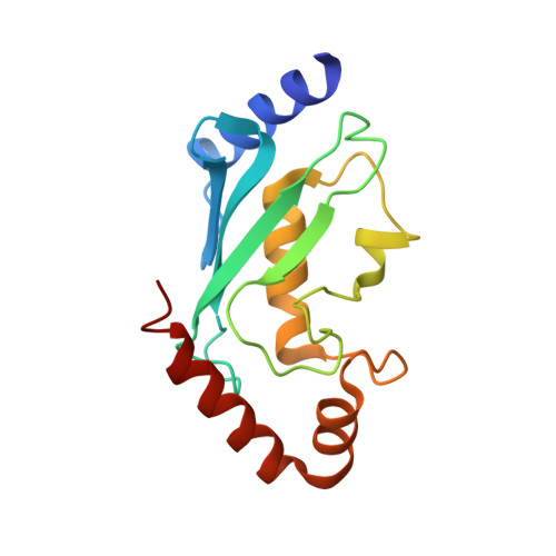
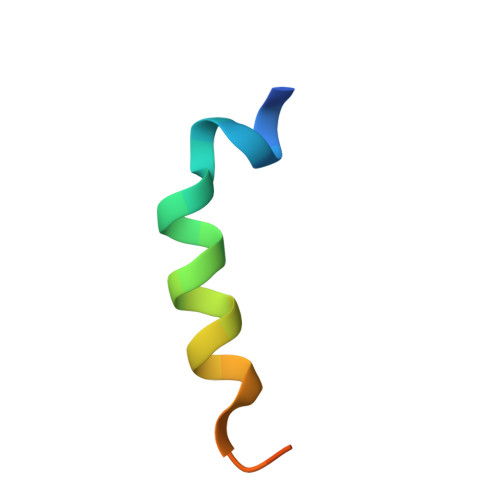
E3 Ligase Rad18 Promotes Monoubiquitination Rather Than Ubiquitin Chain Formation by E2 Enzyme Rad6.

Hibbert, R.G.Huang, A.Boelens, R.Sixma, T.K.

(2011) Proc Natl Acad Sci U S A 108: 5590

  • DOI: https://doi.org/10.1073/pnas.1017516108
  • Primary Citation of Related Structures:  
    2YB6, 2YBF

  • PubMed Abstract: 

    In ubiquitin conjugation, different combinations of E2 and E3 enzymes catalyse either monoubiquitination or ubiquitin chain formation. The E2/E3 complex Rad6/Rad18 exclusively monoubiquitinates the proliferating cell nuclear antigen (PCNA) to signal for "error prone" DNA damage tolerance, whereas a different set of conjugation enzymes is required for ubiquitin chain formation on PCNA. Here we show that human E2 enzyme Rad6b is intrinsically capable of catalyzing ubiquitin chain formation. This activity is prevented during PCNA ubiquitination by the interaction of Rad6 with E3 enzyme Rad18. Using NMR and X-ray crystallography we show that the R6BD of Rad18 inhibits this activity by competing with ubiquitin for a noncovalent "backside" binding site on Rad6. Our findings provide mechanistic insights into how E3 enzymes can regulate the ubiquitin conjugation process.


  • Organizational Affiliation

    Division of Biochemistry and Center for Biomedical Genetics, Netherlands Cancer Institute, Plesmanlaan 121, 1066 CX Amsterdam, The Netherlands.


Macromolecules
Find similar proteins by:  (by identity cutoff)  |  3D Structure
Entity ID: 1
MoleculeChains Sequence LengthOrganismDetailsImage
UBIQUITIN-CONJUGATING ENZYME E2 B152Homo sapiensMutation(s): 0 
EC: 6.3.2.19
UniProt & NIH Common Fund Data Resources
Find proteins for P63146 (Homo sapiens)
Explore P63146 
Go to UniProtKB:  P63146
PHAROS:  P63146
GTEx:  ENSG00000119048 
Entity Groups  
Sequence Clusters30% Identity50% Identity70% Identity90% Identity95% Identity100% Identity
UniProt GroupP63146
Sequence Annotations
Expand
  • Reference Sequence
Find similar proteins by:  (by identity cutoff)  |  3D Structure
Entity ID: 2
MoleculeChains Sequence LengthOrganismDetailsImage
E3 UBIQUITIN-PROTEIN LIGASE RAD1827Homo sapiensMutation(s): 0 
EC: 6.3.2
UniProt & NIH Common Fund Data Resources
Find proteins for Q9NS91 (Homo sapiens)
Explore Q9NS91 
Go to UniProtKB:  Q9NS91
PHAROS:  Q9NS91
GTEx:  ENSG00000070950 
Entity Groups  
Sequence Clusters30% Identity50% Identity70% Identity90% Identity95% Identity100% Identity
UniProt GroupQ9NS91
Sequence Annotations
Expand
  • Reference Sequence
Experimental Data & Validation

Experimental Data

  • Method: X-RAY DIFFRACTION
  • Resolution: 2.00 Å
  • R-Value Free: 0.242 
  • R-Value Work: 0.199 
  • R-Value Observed: 0.201 
  • Space Group: P 65 2 2
Unit Cell:
Length ( Å )Angle ( ˚ )
a = 58.21α = 90
b = 58.21β = 90
c = 167.07γ = 120
Software Package:
Software NamePurpose
REFMACrefinement
XDSdata reduction
SCALAdata scaling
PHASERphasing

Structure Validation

View Full Validation Report

Currently 2YBF does not have a validation slider image.


Entry History 

Deposition Data

Revision History  (Full details and data files)

  • Version 1.0: 2011-04-20
    Type: Initial release
  • Version 1.1: 2011-05-08
    Changes: Version format compliance
  • Version 1.2: 2011-07-13
    Changes: Version format compliance
  • Version 1.3: 2023-12-20
    Changes: Data collection, Database references, Derived calculations, Other, Refinement description