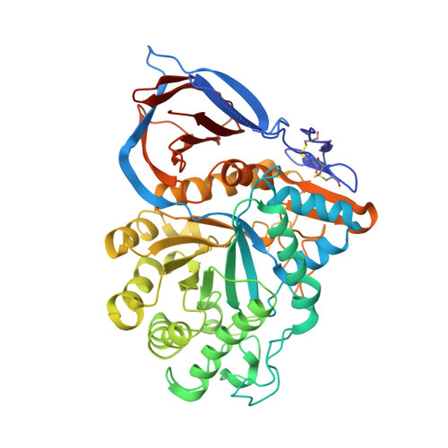
Cyclodextrin-Mediated Crystallization of Acid Beta-Glucosidase in Complex with Amphiphilic Bicyclic Nojirimycin Analogues.
Brumshtein, B., Aguilar-Moncayo, M., Benito, J.M., Garcia Fernandez, J.M., Silman, I., Shaaltiel, Y., Aviezer, D., Sussman, J.L., Futerman, A.H., Ortiz Mellet, C.(2011) Org Biomol Chem 9: 4160
- PubMed: 21483943
- DOI: https://doi.org/10.1039/c1ob05200d
- Primary Citation of Related Structures:
2XWD, 2XWE - PubMed Abstract:
Cyclodextrin-based host-guest chemistry has been exploited to facilitate co-crystallization of recombinant human acid β-glucosidase (β-glucocerebrosidase, GlcCerase) with amphiphilic bicyclic nojirimycin analogues of the sp(2)-iminosugar type. Attempts to co-crystallize GlcCerase with 5-N,6-O-[N'-(n-octyl)iminomethylidene]nojirimycin (NOI-NJ) or with 5-N,6-S-[N'-(n-octyl)iminomethylidene]-6-thionojirimycin (6S-NOI-NJ), two potent inhibitors of the enzyme with promising pharmacological chaperone activity for several Gaucher disease-associated mutations, were unsuccessful probably due to the formation of aggregates that increase the heterogeneity of the sample and affect nucleation and growth of crystals. Cyclomaltoheptaose (β-cyclodextrin, βCD) efficiently captures NOI-NJ and 6S-NOI-NJ in aqueous media to form inclusion complexes in which the lipophilic tail is accommodated in the hydrophobic cavity of the cyclooligosaccharide. The dissociation constant of the complex of the amphiphilic sp(2)-iminosugars with βCD is two orders of magnitude higher than that of the corresponding complex with GlcCerase, allowing the efficient transfer of the inhibitor from the βCD cavity to the GlcCerase active site. Enzyme-inhibitor complexes suitable for X-ray analysis were thus grown in the presence of βCD. In contrast to what was previously observed for the complex of GlcCerase with the more basic derivative, 6-amino-6-deoxy-5-N,6-N-[N'-(n-octyl)iminomethylidene]nojirimycin (6N-NOI-NJ), the β-anomers of both NOI-NJ and 6S-NOI-NJ were seen in the active site, even though the α-anomer was exclusively detected both in aqueous solution and in the corresponding βCD:sp(2)-iminosugar complexes. Our results further suggest that cyclodextrin derivatives might serve as suitable delivery systems of amphiphilic glycosidase inhibitors in a biomedical context.
Organizational Affiliation:
Department of Structural Biology, Weizmann Institute of Science, Rehovot, 76100, Israel.