2WX3

Asymmetric trimer of the human DCP1a C-terminal domain


Experimental Data Snapshot

  • Method: X-RAY DIFFRACTION
  • Resolution: 2.31 Å
  • R-Value Free: 0.252 
  • R-Value Work: 0.208 
  • R-Value Observed: 0.210 

wwPDB Validation   3D Report Full Report


This is version 1.2 of the entry. See complete history


Literature

Dcp1 Forms Asymmetric Trimers to Assemble Into Active Mrna Decapping Complexes in Metazoa.

Tritschler, F.Braun, J.E.Motz, C.Igreja, C.Haas, G.Truffault, V.Izaurralde, E.Weichenrieder, O.

(2009) Proc Natl Acad Sci U S A 106: 21591

  • DOI: https://doi.org/10.1073/pnas.0909871106
  • Primary Citation of Related Structures:  
    2WX3, 2WX4

  • PubMed Abstract: 

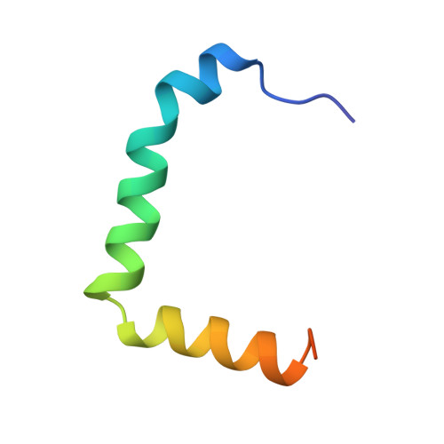
    DCP1 stimulates the decapping enzyme DCP2, which removes the mRNA 5' cap structure committing mRNAs to degradation. In multicellular eukaryotes, DCP1-DCP2 interaction is stabilized by additional proteins, including EDC4. However, most information on DCP2 activation stems from studies in S. cerevisiae, which lacks EDC4. Furthermore, DCP1 orthologs from multicellular eukaryotes have a C-terminal extension, absent in fungi. Here, we show that in metazoa, a conserved DCP1 C-terminal domain drives DCP1 trimerization. Crystal structures of the DCP1-trimerization domain reveal an antiparallel assembly comprised of three kinked alpha-helices. Trimerization is required for DCP1 to be incorporated into active decapping complexes and for efficient mRNA decapping in vivo. Our results reveal an unexpected connectivity and complexity of the mRNA decapping network in multicellular eukaryotes, which likely enhances opportunities for regulating mRNA degradation.


  • Organizational Affiliation

    Department of Biochemistry, Max Planck Institute for Developmental Biology, Spemannstrasse 35, D-72076 Tübingen, Germany.


Macromolecules
Find similar proteins by:  (by identity cutoff)  |  3D Structure
Entity ID: 1
MoleculeChains Sequence LengthOrganismDetailsImage
MRNA-DECAPPING ENZYME 1A
A, B, C
51Homo sapiensMutation(s): 0 
UniProt & NIH Common Fund Data Resources
Find proteins for Q9NPI6 (Homo sapiens)
Explore Q9NPI6 
Go to UniProtKB:  Q9NPI6
PHAROS:  Q9NPI6
GTEx:  ENSG00000272886 
Entity Groups  
Sequence Clusters30% Identity50% Identity70% Identity90% Identity95% Identity100% Identity
UniProt GroupQ9NPI6
Sequence Annotations
Expand
  • Reference Sequence
Experimental Data & Validation

Experimental Data

  • Method: X-RAY DIFFRACTION
  • Resolution: 2.31 Å
  • R-Value Free: 0.252 
  • R-Value Work: 0.208 
  • R-Value Observed: 0.210 
  • Space Group: P 32 2 1
Unit Cell:
Length ( Å )Angle ( ˚ )
a = 64.75α = 90
b = 64.75β = 90
c = 103.03γ = 120
Software Package:
Software NamePurpose
REFMACrefinement
XDSdata reduction
XSCALEdata scaling
autoSHARPphasing

Structure Validation

View Full Validation Report


Entry History 

Deposition Data

Revision History  (Full details and data files)

  • Version 1.0: 2009-12-01
    Type: Initial release
  • Version 1.1: 2011-05-08
    Changes: Version format compliance
  • Version 1.2: 2011-07-13
    Changes: Version format compliance