2V77

Crystal Structure of Human Carboxypeptidase A1


Experimental Data Snapshot

  • Method: X-RAY DIFFRACTION
  • Resolution: 1.60 Å
  • R-Value Free: 0.192 
  • R-Value Work: 0.178 
  • R-Value Observed: 0.178 

wwPDB Validation   3D Report Full Report


Ligand Structure Quality Assessment 


This is version 1.5 of the entry. See complete history


Literature

Direct interaction between a human digestive protease and the mucoadhesive poly(acrylic acid).

Pallares, I.Fernandez, D.Comellas-Bigler, M.Fernandez-Recio, J.Ventura, S.Aviles, F.X.Bode, W.Vendrell, J.

(2008) Acta Crystallogr D Biol Crystallogr D64: 784-791

  • DOI: https://doi.org/10.1107/S0907444908013474
  • Primary Citation of Related Structures:  
    2V77

  • PubMed Abstract: 

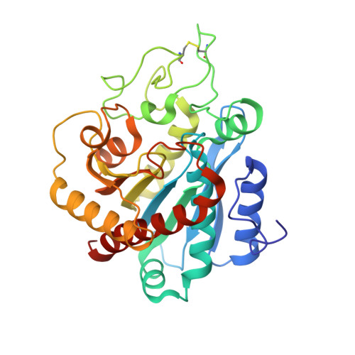
    Carboxypeptidase A1 has been the subject of extensive research in the last 30 y and is one of the most widely studied zinc metalloenzymes. However, the three-dimensional structure of the human form of the enzyme is not yet available. This report describes the three-dimensional structure of human carboxypeptidase A1 (hCPA1) derived from crystals that belong to the tetragonal space group P4(3)2(1)2 and diffract to 1.6 angstroms resolution. A description of the ternary complex hCPA1-Zn2+-poly(acrylic acid) is included as a model of the interaction of mucoadhesive polymers with proteases in the gastrointestinal tract. The direct mode of interaction between poly(acrylic acid) and the active site of the target protease was confirmed by in vitro inhibition assays. The structure was further analyzed in silico through the optimal docking-area method. The characterization of binding sites on the surface of hCPA1 and a comparison with other available carboxypeptidase structures provided further insights into the formation of multiprotein complexes and the activation mechanisms of carboxypeptidase zymogens. The high-resolution structure of hCPA1 provides an excellent template for the modelling of physiologically relevant carboxypeptidases and could also contribute to the design of specific agents for biomedical purposes.


  • Organizational Affiliation

    Departament de Bioquímica i Biologia Molecular, Facultat de Biociències and Institut de Biotecnologia i de Biomedicina, Universitat Autònoma de Barcelona, E-08193 Bellaterra, Spain.


Macromolecules
Find similar proteins by:  (by identity cutoff)  |  3D Structure
Entity ID: 1
MoleculeChains Sequence LengthOrganismDetailsImage
CARBOXYPEPTIDASE A1
A, B
309Homo sapiensMutation(s): 0 
UniProt & NIH Common Fund Data Resources
Find proteins for P15085 (Homo sapiens)
Explore P15085 
Go to UniProtKB:  P15085
PHAROS:  P15085
GTEx:  ENSG00000091704 
Entity Groups  
Sequence Clusters30% Identity50% Identity70% Identity90% Identity95% Identity100% Identity
UniProt GroupP15085
Sequence Annotations
Expand
  • Reference Sequence
Small Molecules
Binding Affinity Annotations 
IDSourceBinding Affinity
PAY Binding MOAD:  2V77 Ki: 8700 (nM) from 1 assay(s)
Experimental Data & Validation

Experimental Data

  • Method: X-RAY DIFFRACTION
  • Resolution: 1.60 Å
  • R-Value Free: 0.192 
  • R-Value Work: 0.178 
  • R-Value Observed: 0.178 
  • Space Group: P 43 21 2
Unit Cell:
Length ( Å )Angle ( ˚ )
a = 129.565α = 90
b = 129.565β = 90
c = 90.856γ = 90
Software Package:
Software NamePurpose
CNSrefinement
DENZOdata reduction
SCALEPACKdata scaling
AMoREphasing

Structure Validation

View Full Validation Report


Ligand Structure Quality Assessment 


Entry History 

Deposition Data

Revision History  (Full details and data files)

  • Version 1.0: 2008-07-01
    Type: Initial release
  • Version 1.1: 2011-05-08
    Changes: Version format compliance
  • Version 1.2: 2011-07-13
    Changes: Version format compliance
  • Version 1.3: 2017-07-19
    Changes: Database references
  • Version 1.4: 2019-05-22
    Changes: Data collection, Refinement description
  • Version 1.5: 2023-12-13
    Changes: Advisory, Data collection, Database references, Derived calculations, Other, Refinement description