2UWN

Crystal structure of Human Complement Factor H, SCR domains 6-8 (H402 risk variant), in complex with ligand.


Experimental Data Snapshot

  • Method: X-RAY DIFFRACTION
  • Resolution: 2.35 Å
  • R-Value Free: 0.246 
  • R-Value Work: 0.214 
  • R-Value Observed: 0.214 

wwPDB Validation   3D Report Full Report


This is version 2.1 of the entry. See complete history


Literature

Structural Basis for Complement Factor H Linked Age-Related Macular Degeneration.

Prosser, B.E.Johnson, S.Roversi, P.Herbert, A.P.Blaum, B.S.Tyrrell, J.Jowitt, T.A.Clark, S.J.Tarelli, E.Uhrin, D.Barlow, P.N.Sim, R.B.Day, A.J.Lea, S.M.

(2007) J Exp Med 204: 2277

  • DOI: https://doi.org/10.1084/jem.20071069
  • Primary Citation of Related Structures:  
    2UWN, 2V8E

  • PubMed Abstract: 

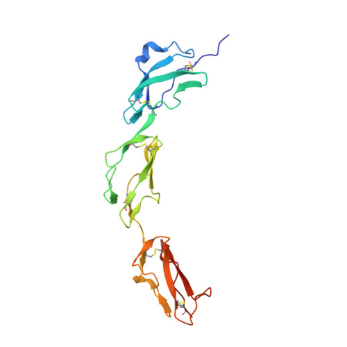
    Nearly 50 million people worldwide suffer from age-related macular degeneration (AMD), which causes severe loss of central vision. A single-nucleotide polymorphism in the gene for the complement regulator factor H (FH), which causes a Tyr-to-His substitution at position 402, is linked to approximately 50% of attributable risks for AMD. We present the crystal structure of the region of FH containing the polymorphic amino acid His402 in complex with an analogue of the glycosaminoglycans (GAGs) that localize the complement regulator on the cell surface. The structure demonstrates direct coordination of ligand by the disease-associated polymorphic residue, providing a molecular explanation of the genetic observation. This glycan-binding site occupies the center of an extended interaction groove on the regulator's surface, implying multivalent binding of sulfated GAGs. This finding is confirmed by structure-based site-directed mutagenesis, nuclear magnetic resonance-monitored binding experiments performed for both H402 and Y402 variants with this and another model GAG, and analysis of an extended GAG-FH complex.


  • Organizational Affiliation

    Sir William Dunn School of Pathology, University of Oxford, Oxford, OX1 3RE, England, UK.


Macromolecules
Find similar proteins by:  (by identity cutoff)  |  3D Structure
Entity ID: 1
MoleculeChains Sequence LengthOrganismDetailsImage
HUMAN COMPLEMENT FACTOR H187Homo sapiensMutation(s): 0 
UniProt & NIH Common Fund Data Resources
Find proteins for P08603 (Homo sapiens)
Explore P08603 
Go to UniProtKB:  P08603
PHAROS:  P08603
GTEx:  ENSG00000000971 
Entity Groups  
Sequence Clusters30% Identity50% Identity70% Identity90% Identity95% Identity100% Identity
UniProt GroupP08603
Sequence Annotations
Expand
  • Reference Sequence
Oligosaccharides

Help

Entity ID: 2
MoleculeChains Length2D Diagram Glycosylation3D Interactions
1,3,4,6-tetra-O-sulfo-beta-D-fructofuranose-(2-1)-2,3,4,6-tetra-O-sulfonato-alpha-D-glucopyranose
B
2N/A
Glycosylation Resources
GlyTouCan:  G97555GV
GlyCosmos:  G97555GV
Biologically Interesting Molecules (External Reference) 1 Unique
Experimental Data & Validation

Experimental Data

  • Method: X-RAY DIFFRACTION
  • Resolution: 2.35 Å
  • R-Value Free: 0.246 
  • R-Value Work: 0.214 
  • R-Value Observed: 0.214 
  • Space Group: C 2 2 21
Unit Cell:
Length ( Å )Angle ( ˚ )
a = 74.756α = 90
b = 92.491β = 90
c = 57.311γ = 90
Software Package:
Software NamePurpose
DMmodel building
MOSFLMdata reduction
SCALAdata scaling
MOLREPphasing
BUSTER-TNTphasing
SHARP-SOLOMONphasing
DMphasing
piratephasing

Structure Validation

View Full Validation Report


Entry History 

Revision History  (Full details and data files)

  • Version 1.0: 2007-10-02
    Type: Initial release
  • Version 1.1: 2011-05-08
    Changes: Version format compliance
  • Version 1.2: 2011-07-13
    Changes: Version format compliance
  • Version 2.0: 2020-07-29
    Type: Remediation
    Reason: Carbohydrate remediation
    Changes: Advisory, Atomic model, Data collection, Derived calculations, Non-polymer description, Other, Refinement description, Structure summary
  • Version 2.1: 2023-12-13
    Changes: Data collection, Database references, Refinement description, Structure summary