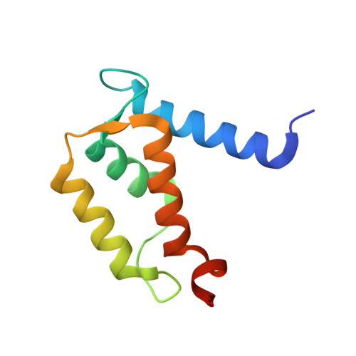
2RGI

Crystal structure of Ca2+-free S100A2 at 1.6 A resolution


Experimental Data Snapshot

  • Method: X-RAY DIFFRACTION
  • Resolution: 1.60 Å
  • R-Value Free: 0.227 
  • R-Value Work: 0.186 
  • R-Value Observed: 0.188 

wwPDB Validation   3D Report Full Report


This is version 1.3 of the entry. See complete history


Literature

Crystal structure of Ca2+ -free S100A2 at 1.6-A resolution.

Koch, M.Diez, J.Fritz, G.

(2008) J Mol Biol 378: 931-940

  • DOI: https://doi.org/10.1016/j.jmb.2008.03.019
  • Primary Citation of Related Structures:  
    2RGI

  • PubMed Abstract: 

    S100A2 is an EF hand-containing Ca(2+)-binding protein of the family of S100 proteins. The protein is localized exclusively in the nucleus and is involved in cell cycle regulation. It attracted most interest by its function as a tumor suppressor via p53 interaction. We determined the crystal structure of homodimeric S100A2 in the Ca(2+)-free state at 1.6-A resolution. The structure revealed structural differences between subunits A and B, especially in the conformation of a loop that connects the N- and C-terminal EF hands and represents a part of the target-binding site in S100 proteins. Analysis of the hydrogen bonding network and molecular dynamics calculations indicate that one of the two observed conformations is more stable. The structure revealed Na(+) bound to each N-terminal EF hand of both subunits coordinated by oxygen atoms of the backbone carbonyl and water molecules. Comparison with the structures of Ca(2+)-free S100A3 and S100A6 suggests that Na(+) might occupy the S100-specific EF hand in the Ca(2+)-free state.


  • Organizational Affiliation

    Fachbereich Biologie, Universität Konstanz, Postfach M665, Universitätsstrasse 10, 78457 Konstanz, Germany.


Macromolecules
Find similar proteins by:  (by identity cutoff)  |  3D Structure
Entity ID: 1
MoleculeChains Sequence LengthOrganismDetailsImage
Protein S100-A2
A, B
98Homo sapiensMutation(s): 4 
Gene Names: S100A2S100L
UniProt & NIH Common Fund Data Resources
Find proteins for P29034 (Homo sapiens)
Explore P29034 
Go to UniProtKB:  P29034
PHAROS:  P29034
GTEx:  ENSG00000196754 
Entity Groups  
Sequence Clusters30% Identity50% Identity70% Identity90% Identity95% Identity100% Identity
UniProt GroupP29034
Sequence Annotations
Expand
  • Reference Sequence
Experimental Data & Validation

Experimental Data

  • Method: X-RAY DIFFRACTION
  • Resolution: 1.60 Å
  • R-Value Free: 0.227 
  • R-Value Work: 0.186 
  • R-Value Observed: 0.188 
  • Space Group: P 21 21 21
Unit Cell:
Length ( Å )Angle ( ˚ )
a = 43.516α = 90
b = 57.82β = 90
c = 59.794γ = 90
Software Package:
Software NamePurpose
XSCALEdata scaling
PHASERphasing
REFMACrefinement
PDB_EXTRACTdata extraction
XDSdata reduction

Structure Validation

View Full Validation Report


Entry History 

Deposition Data

Revision History  (Full details and data files)

  • Version 1.0: 2008-01-29
    Type: Initial release
  • Version 1.1: 2011-07-13
    Changes: Version format compliance
  • Version 1.2: 2021-10-20
    Changes: Database references, Derived calculations
  • Version 1.3: 2023-08-30
    Changes: Data collection, Refinement description