2Q7O

Structure of human purine nucleoside phosphorylase in complex with L-Immucillin-H


Experimental Data Snapshot

  • Method: X-RAY DIFFRACTION
  • Resolution: 2.90 Å
  • R-Value Free: 0.262 
  • R-Value Work: 0.216 
  • R-Value Observed: 0.218 

wwPDB Validation   3D Report Full Report


Ligand Structure Quality Assessment 


This is version 1.3 of the entry. See complete history


Literature

L-Enantiomers of transition state analogue inhibitors bound to human purine nucleoside phosphorylase.

Rinaldo-Matthis, A.Murkin, A.S.Ramagopal, U.A.Clinch, K.Mee, S.P.Evans, G.B.Tyler, P.C.Furneaux, R.H.Almo, S.C.Schramm, V.L.

(2008) J Am Chem Soc 130: 842-844

  • DOI: https://doi.org/10.1021/ja710733g
  • Primary Citation of Related Structures:  
    2Q7O, 3BGS

  • PubMed Abstract: 

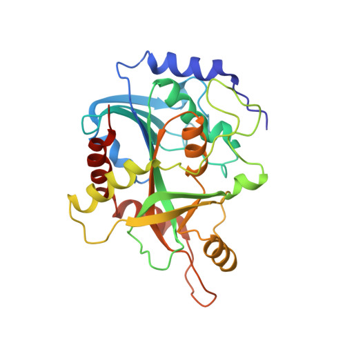
    Human purine nucleoside phosphorylase (PNP) was crystallized with transition-state analogue inhibitors Immucillin-H and DADMe-Immucillin-H synthesized with ribosyl mimics of l-stereochemistry. The inhibitors demonstrate that major driving forces for tight binding of these analogues are the leaving group interaction and the cationic mimicry of the transition state, even though large geometric changes occur with d-Immucillins and l-Immucillins bound to human PNP.


  • Organizational Affiliation

    Department of Biochemistry, Albert Einstein College of Medicine, Bronx, New York 10461, USA.


Macromolecules
Find similar proteins by:  (by identity cutoff)  |  3D Structure
Entity ID: 1
MoleculeChains Sequence LengthOrganismDetailsImage
Purine nucleoside phosphorylaseA [auth E]289Homo sapiensMutation(s): 0 
Gene Names: NPPNP
EC: 2.4.2.1
UniProt & NIH Common Fund Data Resources
Find proteins for P00491 (Homo sapiens)
Explore P00491 
Go to UniProtKB:  P00491
PHAROS:  P00491
GTEx:  ENSG00000198805 
Entity Groups  
Sequence Clusters30% Identity50% Identity70% Identity90% Identity95% Identity100% Identity
UniProt GroupP00491
Sequence Annotations
Expand
  • Reference Sequence
Small Molecules
Ligands 2 Unique
IDChains Name / Formula / InChI Key2D Diagram3D Interactions
IMH
Query on IMH

Download Ideal Coordinates CCD File 
C [auth E]1,4-DIDEOXY-4-AZA-1-(S)-(9-DEAZAHYPOXANTHIN-9-YL)-D-RIBITOL
C11 H14 N4 O4
IWKXDMQDITUYRK-KUBHLMPHSA-N
PO4
Query on PO4

Download Ideal Coordinates CCD File 
B [auth E]PHOSPHATE ION
O4 P
NBIIXXVUZAFLBC-UHFFFAOYSA-K
Binding Affinity Annotations 
IDSourceBinding Affinity
IMH PDBBind:  2Q7O Ki: 12 (nM) from 1 assay(s)
Experimental Data & Validation

Experimental Data

  • Method: X-RAY DIFFRACTION
  • Resolution: 2.90 Å
  • R-Value Free: 0.262 
  • R-Value Work: 0.216 
  • R-Value Observed: 0.218 
  • Space Group: H 3 2
Unit Cell:
Length ( Å )Angle ( ˚ )
a = 143.457α = 90
b = 143.457β = 90
c = 166.248γ = 120
Software Package:
Software NamePurpose
DENZOdata reduction
SCALEPACKdata scaling
MOLREPphasing
REFMACrefinement
PDB_EXTRACTdata extraction
CBASSdata collection
HKL-2000data reduction

Structure Validation

View Full Validation Report


Ligand Structure Quality Assessment 


Entry History 

Deposition Data

Revision History  (Full details and data files)

  • Version 1.0: 2008-01-15
    Type: Initial release
  • Version 1.1: 2011-07-13
    Changes: Derived calculations, Version format compliance
  • Version 1.2: 2017-10-18
    Changes: Refinement description
  • Version 1.3: 2023-08-30
    Changes: Data collection, Database references, Derived calculations, Refinement description, Structure summary