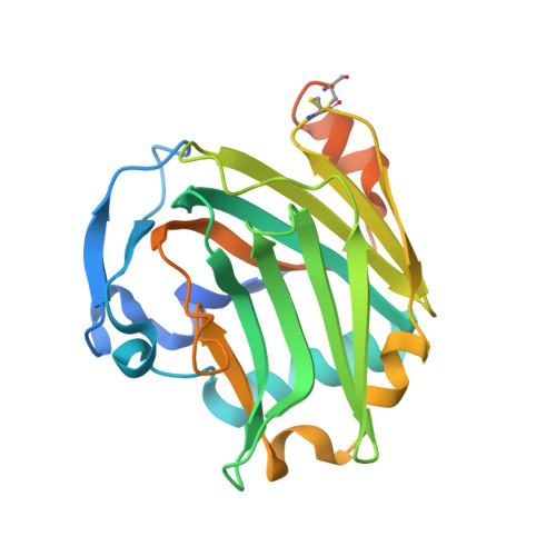
Heparin-induced cis- and trans-Dimerization Modes of the Thrombospondin-1 N-terminal Domain.
Tan, K., Duquette, M., Liu, J.H., Shanmugasundaram, K., Joachimiak, A., Gallagher, J.T., Rigby, A.C., Wang, J.H., Lawler, J.(2008) J Biol Chem 283: 3932-3941
- PubMed: 18065761
- DOI: https://doi.org/10.1074/jbc.M705203200
- Primary Citation of Related Structures:
2ES3, 2OUH, 2OUJ - PubMed Abstract:
Through its interactions with proteins and proteoglycans, thrombospondin-1 (TSP-1) functions at the interface of the cell membrane and the extracellular matrix to regulate matrix structure and cellular phenotype. We have previously determined the structure of the high affinity heparin-binding domain of TSP-1, designated TSPN-1, in association with the synthetic heparin, Arixtra. To establish that the binding of TSPN-1 to Arixtra is representative of the association with naturally occurring heparins, we have determined the structures of TSPN-1 in complex with heparin oligosaccharides containing eight (dp8) and ten (dp10) subunits, by x-ray crystallography. We have found that dp8 and dp10 bind to TSPN-1 in a manner similar to Arixtra and that dp8 and dp10 induce the formation of trans and cis TSPN-1 dimers, respectively. In silico docking calculations partnered with our crystal structures support the importance of arginine residues in positions 29, 42, and 77 in binding sulfate groups of the dp8 and dp10 forms of heparin. The ability of several TSPN-1 domains to bind to glycosaminoglycans simultaneously probably increases the affinity of binding through multivalent interactions. The formation of cis and trans dimers of the TSPN-1 domain with relatively short segments of heparin further enhances the ability of TSP-1 to participate in high affinity binding to glycosaminoglycans. Dimer formation may also involve TSPN-1 domains from two separate TSP-1 molecules. This association would enable glycosaminoglycans to cluster TSP-1.
Organizational Affiliation:
Department of Medical Oncology, Dana-Farber Cancer Institute, Boston, Massachusetts 02115, USA.