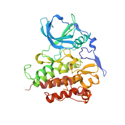
The 2.7 A crystal structure of the autoinhibited human c-Fms kinase domain.
Walter, M., Lucet, I.S., Patel, O., Broughton, S.E., Bamert, R., Williams, N.K., Fantino, E., Wilks, A.F., Rossjohn, J.(2007) J Mol Biol 367: 839-847
- PubMed: 17292918
- DOI: https://doi.org/10.1016/j.jmb.2007.01.036
- Primary Citation of Related Structures:
2OGV - PubMed Abstract:
c-Fms, a member of the Platelet-derived Growth Factor (PDGF) receptor family of receptor tyrosine kinases (RTKs), is the receptor for macrophage colony stimulating factor (CSF-1) that regulates proliferation, differentiation and survival of cells of the mononuclear phagocyte lineage. Abnormal expression of c-fms proto-oncogene is associated with a significant number of human pathologies, including a variety of cancers and rheumatoid arthritis. Accordingly, c-Fms represents an attractive therapeutic target. To further understand the regulation of c-Fms, we determined the 2.7 A resolution crystal structure of the cytosolic domain of c-Fms that comprised the kinase domain and the juxtamembrane domain. The structure reveals the crucial inhibitory role of the juxtamembrane domain (JM) that binds to a hydrophobic site immediately adjacent to the ATP binding pocket. This interaction prevents the activation loop from adopting an active conformation thereby locking the c-Fms kinase into an autoinhibited state. As observed for other members of the PDGF receptor family, namely c-Kit and Flt3, three JM-derived tyrosine residues primarily drive the mechanism for autoinhibition in c-Fms, therefore defining a common autoinhibitory mechanism within this family. Moreover the structure provides an understanding of c-Fms inhibition by Gleevec as well as providing a platform for the development of more selective inhibitors that target the inactive conformation of c-Fms kinase.
Organizational Affiliation:
Protein Crystallography Unit, Department of Biochemistry and Molecular Biology, School of Biomedical Sciences, Monash University, Clayton, Victoria 3800, Australia.