2O9I

Crystal Structure of the Human Pregnane X Receptor LBD in complex with an SRC-1 coactivator peptide and T0901317


Experimental Data Snapshot

  • Method: X-RAY DIFFRACTION
  • Resolution: 2.80 Å
  • R-Value Free: 0.240 
  • R-Value Work: 0.228 
  • R-Value Observed: 0.228 

wwPDB Validation   3D Report Full Report


Ligand Structure Quality Assessment 


This is version 1.4 of the entry. See complete history


Literature

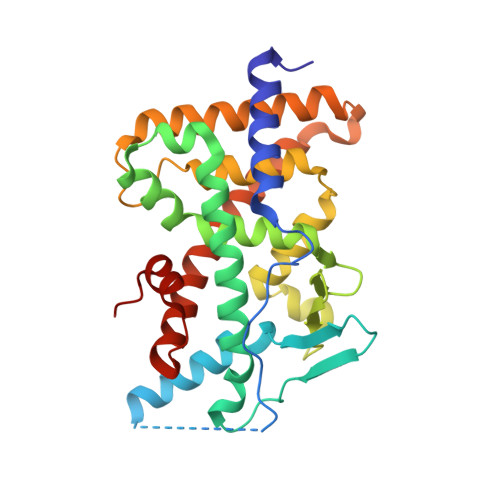
Crystal structure of the PXR-T1317 complex provides a scaffold to examine the potential for receptor antagonism.

Xue, Y.Chao, E.Zuercher, W.J.Willson, T.M.Collins, J.L.Redinbo, M.R.

(2007) Bioorg Med Chem 15: 2156-2166

  • DOI: https://doi.org/10.1016/j.bmc.2006.12.026
  • Primary Citation of Related Structures:  
    2O9I

  • PubMed Abstract: 

    The human pregnane X receptor (PXR) recognizes a range of structurally and chemically distinct ligands and plays a key role in regulating the expression of protective gene products involved in the metabolism and excretion of potentially harmful compounds. The identification and development of PXR antagonists is desirable as a potential way to control the up-regulation of drug metabolism pathways during the therapeutic treatment of disease. We present the 2.8A resolution crystal structure of the PXR ligand binding domain (LBD) in complex with T0901317 (T1317), which is also an agonist of another member of the orphan class of the nuclear receptor superfamily, the liver X receptor (LXR). In spite of differences in the size and shape of the receptors' ligand binding pockets, key interactions with this ligand are conserved between human PXR and human LXR. Based on the PXR-T1317 structure, analogues of T1317 were generated with the goal of designing an PXR antagonist effective via the receptor's ligand binding pocket. We find that selectivity in activating PXR versus LXR was achieved; such compounds may be useful in addressing neurodegenerative diseases like Niemann-Pick C. We were not successful, however, in producing a PXR antagonist. Based on these observations, we conclude that the generation of PXR antagonists targeted to the ligand binding pocket may be difficult due to the promiscuity and structural conformability of this xenobiotic sensor.


  • Organizational Affiliation

    Department of Chemistry, CB#3290, University of North Carolina at Chapel Hill, Chapel Hill, NC 27599-3290, USA.


Macromolecules
Find similar proteins by:  (by identity cutoff)  |  3D Structure
Entity ID: 1
MoleculeChains Sequence LengthOrganismDetailsImage
Orphan nuclear receptor PXR
A, B
293Homo sapiensMutation(s): 1 
Gene Names: NR1I2PXR
UniProt & NIH Common Fund Data Resources
Find proteins for O75469 (Homo sapiens)
Explore O75469 
Go to UniProtKB:  O75469
PHAROS:  O75469
GTEx:  ENSG00000144852 
Entity Groups  
Sequence Clusters30% Identity50% Identity70% Identity90% Identity95% Identity100% Identity
UniProt GroupO75469
Sequence Annotations
Expand
  • Reference Sequence

Find similar proteins by:  Sequence   |   3D Structure  

Entity ID: 2
MoleculeChains Sequence LengthOrganismDetailsImage
Nuclear Receptor Coactivator 1 isoform 3
C, D
15N/AMutation(s): 0 
UniProt & NIH Common Fund Data Resources
Find proteins for P70365 (Mus musculus)
Explore P70365 
Go to UniProtKB:  P70365
IMPC:  MGI:1276523
Entity Groups  
Sequence Clusters30% Identity50% Identity70% Identity90% Identity95% Identity100% Identity
UniProt GroupP70365
Sequence Annotations
Expand
  • Reference Sequence
Small Molecules
Ligands 1 Unique
IDChains Name / Formula / InChI Key2D Diagram3D Interactions
444
Query on 444

Download Ideal Coordinates CCD File 
E [auth A],
F [auth B]
N-(2,2,2-TRIFLUOROETHYL)-N-{4-[2,2,2-TRIFLUORO-1-HYDROXY-1-(TRIFLUOROMETHYL)ETHYL]PHENYL}BENZENESULFONAMIDE
C17 H12 F9 N O3 S
SGIWFELWJPNFDH-UHFFFAOYSA-N
Binding Affinity Annotations 
IDSourceBinding Affinity
444 BindingDB:  2O9I IC50: 40 (nM) from 1 assay(s)
EC50: min: 8, max: 270 (nM) from 7 assay(s)
PDBBind:  2O9I IC50: 40 (nM) from 1 assay(s)
Experimental Data & Validation

Experimental Data

  • Method: X-RAY DIFFRACTION
  • Resolution: 2.80 Å
  • R-Value Free: 0.240 
  • R-Value Work: 0.228 
  • R-Value Observed: 0.228 
  • Space Group: P 21 21 21
Unit Cell:
Length ( Å )Angle ( ˚ )
a = 83.979α = 90
b = 90.618β = 90
c = 105.725γ = 90
Software Package:
Software NamePurpose
CNSrefinement
CrystalCleardata collection
HKL-2000data reduction
HKL-2000data scaling
AMoREphasing

Structure Validation

View Full Validation Report


Ligand Structure Quality Assessment 


Entry History 

Deposition Data

Revision History  (Full details and data files)

  • Version 1.0: 2007-01-30
    Type: Initial release
  • Version 1.1: 2008-05-01
    Changes: Version format compliance
  • Version 1.2: 2011-07-13
    Changes: Version format compliance
  • Version 1.3: 2021-10-20
    Changes: Database references, Derived calculations
  • Version 1.4: 2023-08-30
    Changes: Data collection, Refinement description